The PABPN1 Monoclonal Antibody (CAB1735) is a high-quality antibody developed for reliable detection and analysis of target proteins. This antibody, generated in rabbits, shows high reactivity with human samples and is validated for use in various applications, including Western blot and immunofluorescence.PABPN1, also known as poly(A) binding protein nuclear 1, plays a crucial role in RNA processing and stability, particularly in the regulation of polyadenylation. Dysregulation of PABPN1 has been implicated in diseases such as oculopharyngeal muscular dystrophy (OPMD), highlighting its importance in physiological processes and disease development.
This antibody is validated for use in WB, IHC-P, IF/ICC, ELISA, IF-P applications and has demonstrated reactivity against Human, Mouse, Rat samples.
Product Name:
PABPN1 Monoclonal Antibody
SKU:
CAB1735
Size:
20μL, 100μL
Reactivity:
Human, Mouse, Rat
Clone Number:
ARC0730
Conjugate:
Unconjugated
Immunogen:
Synthetic peptide. This information is considered to be commercially sensitive.
Recommended starting concentration is 1 μg/mL. Please optimize the concentration based on your specific assay requirements.
Synonyms:
OPMD, PAB2, PABII, PABP2, PABP-2, PABPN1
Positive Sample:
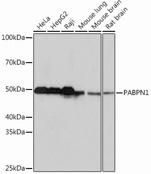
HeLa, HepG2, Raji, Mouse lung, Mouse brain, Rat brain
Cellular Localization:
Cytoplasm, Nucleus.
Calculated MW:
31kDa/33kDa
Observed MW:
50kDa
This gene encodes an abundant nuclear protein that binds with high affinity to nascent poly(A) tails. The protein is required for progressive and efficient polymerization of poly(A) tails at the 3' ends of eukaryotic transcripts and controls the size of the poly(A) tail to about 250 nt. At steady-state, this protein is localized in the nucleus whereas a different poly(A) binding protein is localized in the cytoplasm. This gene contains a GCG trinucleotide repeat at the 5' end of the coding region, and expansion of this repeat from the normal 6 copies to 8-13 copies leads to autosomal dominant oculopharyngeal muscular dystrophy (OPMD) disease. Related pseudogenes have been identified on chromosomes 19 and X. Read-through transcription also exists between this gene and the neighboring upstream BCL2-like 2 (BCL2L2) gene.
Purification Method
Affinity purification
Gene ID
8106
RRID
AB_2861734
Buffer Information
Store at -20℃. Avoid freeze / thaw cycles. Buffer: PBS containing 50% glycerol and 0.05% BSA, preserved with proclin300 or sodium azide, pH 7.3.
Western blot analysis of various lysates using PABPN1 Rabbit mAb (CAB1735) at 1:1000 dilution. Secondary antibody: HRP-conjugated Goat anti-Rabbit IgG (H+L) (CABS014) at 1:10000 dilution. Lysates/proteins: 25μg per lane. Blocking buffer: 3% nonfat dry milk in TBST. Detection: ECL Basic Kit (AbGn00020). Exposure time: 10s.
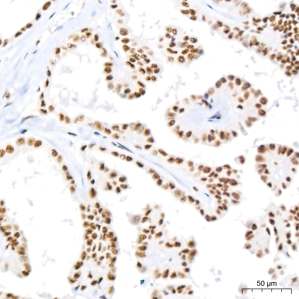
Immunohistochemistry analysis of paraffin-embedded Human cervix cancer tissue using PABPN1 Rabbit mAb (CAB1735) at a dilution of 1:200 (40x lens). High pressure antigen retrieval performed with 0.01M Citrate buffer (pH 6.0) prior to IHC staining.
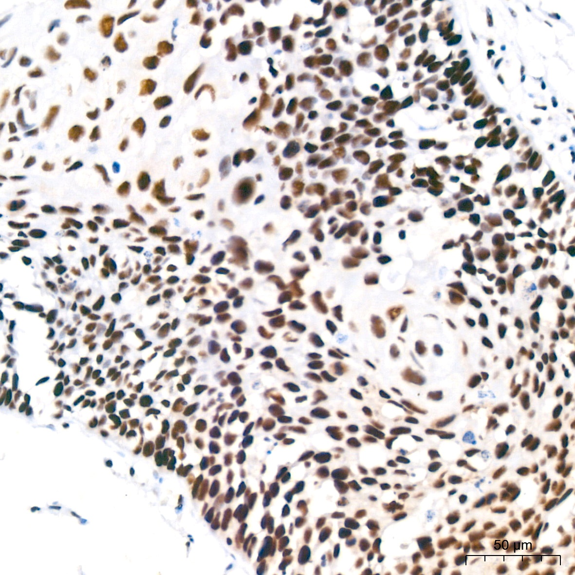
Immunohistochemistry analysis of paraffin-embedded Human lung squamous carcinoma tissue tissue using PABPN1 Rabbit mAb (CAB1735) at a dilution of 1:200 (40x lens). High pressure antigen retrieval performed with 0.01M Citrate buffer (pH 6.0) prior to IHC staining.
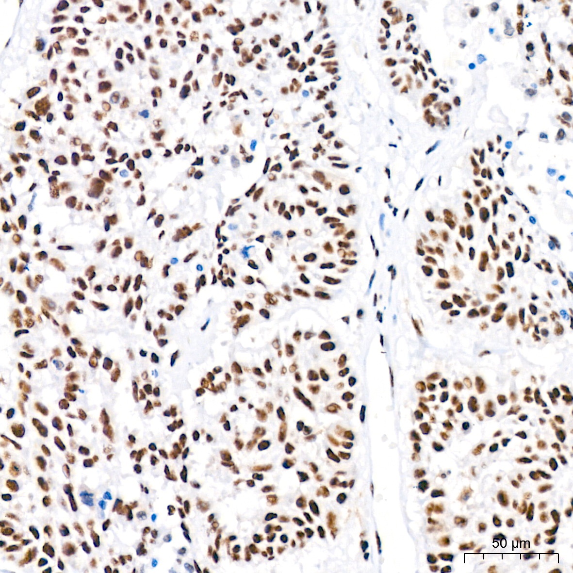
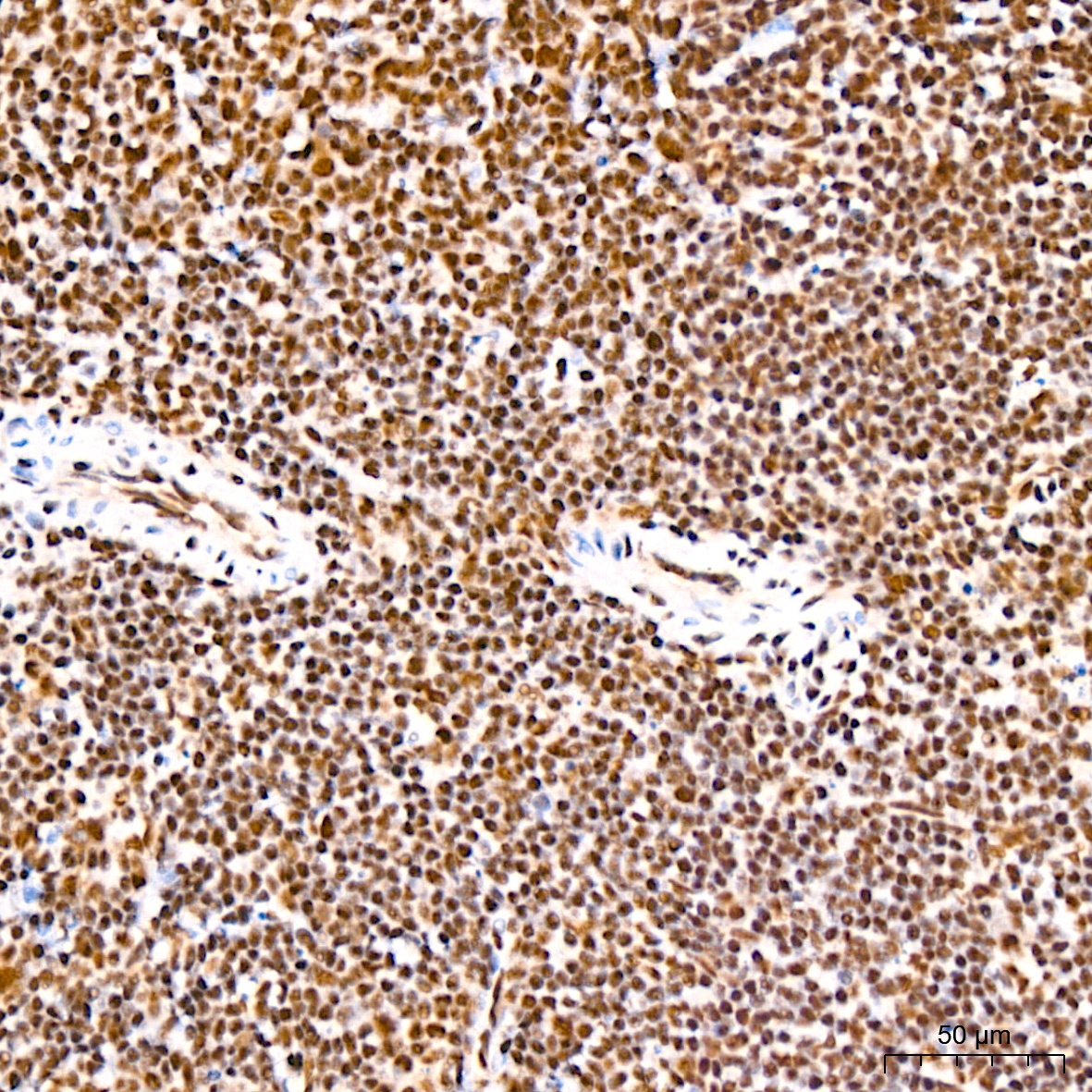
Immunohistochemistry analysis of paraffin-embedded Human thyroid cancer tissue using PABPN1 Rabbit mAb (CAB1735) at a dilution of 1:200 (40x lens). High pressure antigen retrieval performed with 0.01M Citrate buffer (pH 6.0) prior to IHC staining.
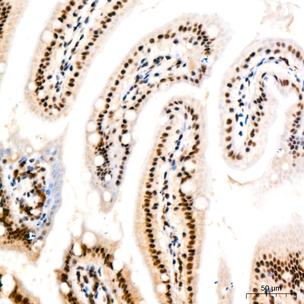
Immunohistochemistry analysis of paraffin-embedded Mouse intestin tissue using PABPN1 Rabbit mAb (CAB1735) at a dilution of 1:200 (40x lens). High pressure antigen retrieval performed with 0.01M Citrate buffer (pH 6.0) prior to IHC staining.
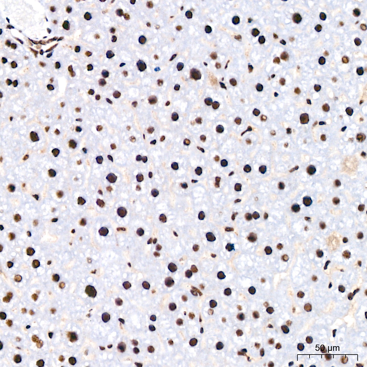
Immunohistochemistry analysis of paraffin-embedded Mouse liver tissue using PABPN1 Rabbit mAb (CAB1735) at a dilution of 1:200 (40x lens). High pressure antigen retrieval performed with 0.01M Citrate buffer (pH 6.0) prior to IHC staining.
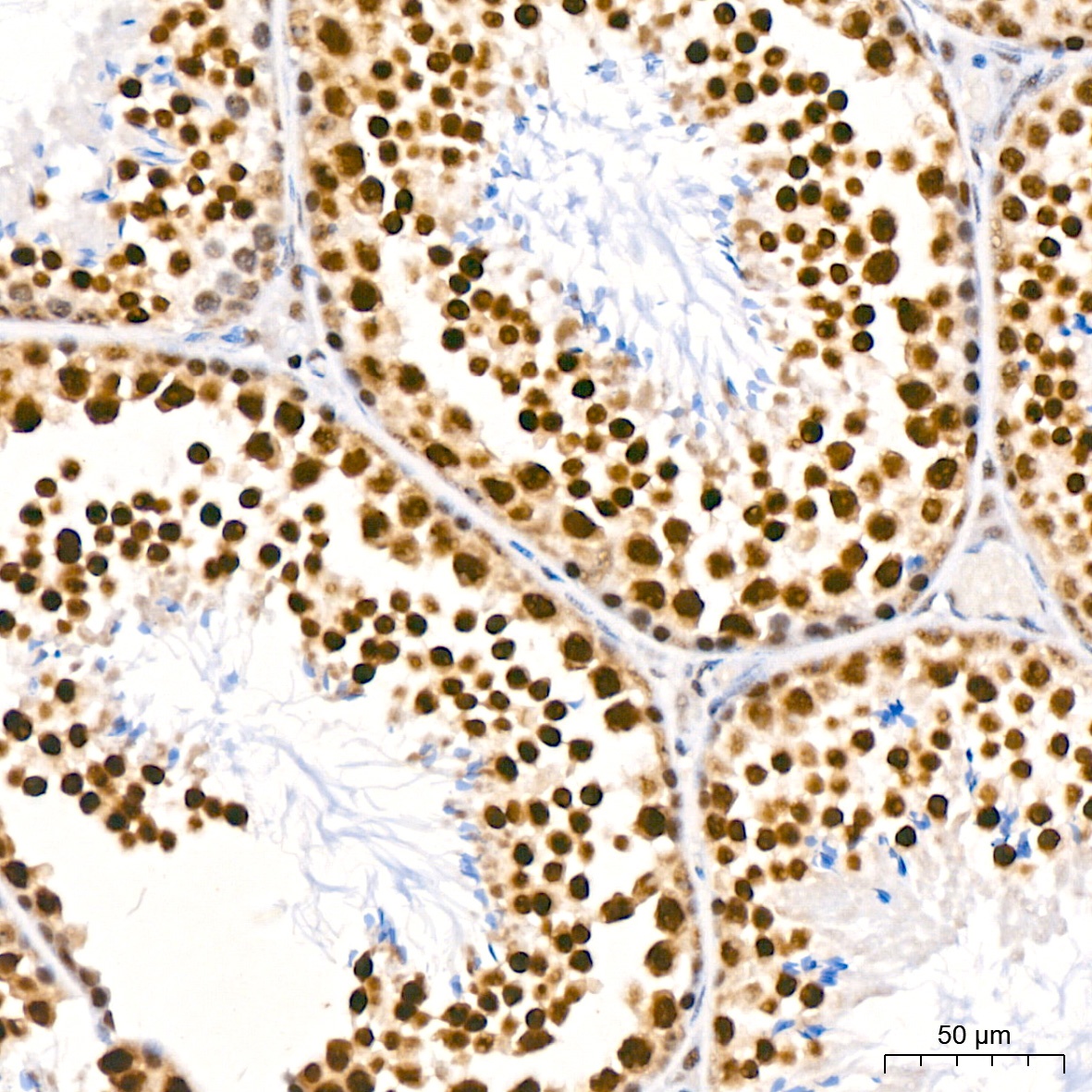
Immunohistochemistry analysis of paraffin-embedded Mouse testis tissue using PABPN1 Rabbit mAb (CAB1735) at a dilution of 1:200 (40x lens). High pressure antigen retrieval performed with 0.01M Citrate buffer (pH 6.0) prior to IHC staining.
Immunohistochemistry analysis of paraffin-embedded Mouse testis tissue using PABPN1 Rabbit mAb (CAB1735) at a dilution of 1:200 (40x lens). High pressure antigen retrieval performed with 0.01M Citrate buffer (pH 6.0) prior to IHC staining.
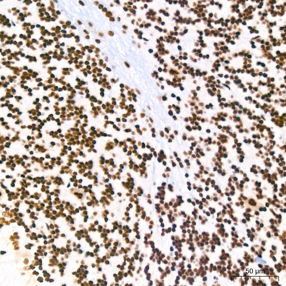
Immunohistochemistry analysis of paraffin-embedded Rat brain tissue using PABPN1 Rabbit mAb (CAB1735) at a dilution of 1:200 (40x lens). High pressure antigen retrieval performed with 0.01M Citrate buffer (pH 6.0) prior to IHC staining.
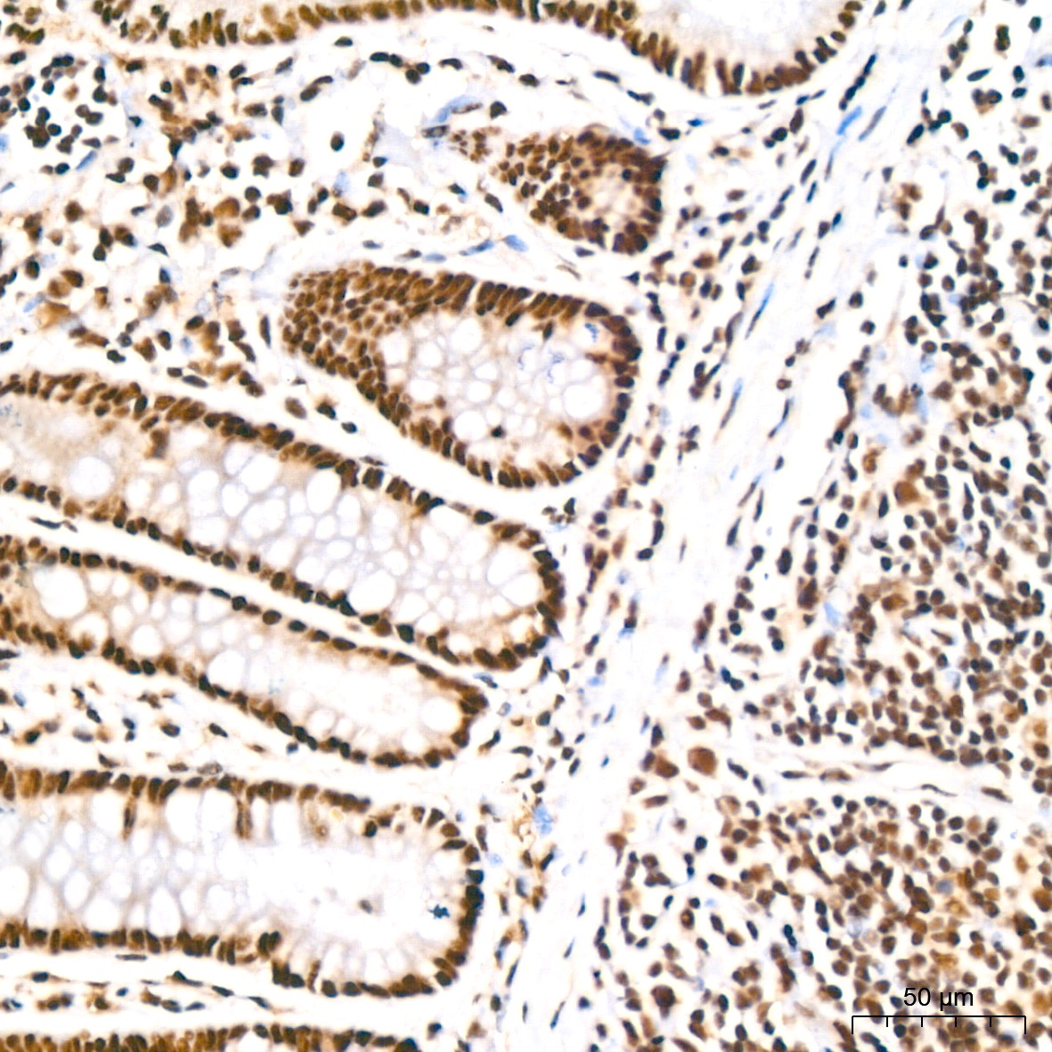
Immunohistochemistry analysis of paraffin-embedded Rat colon tissue using PABPN1 Rabbit mAb (CAB1735) at a dilution of 1:200 (40x lens). High pressure antigen retrieval performed with 0.01M Citrate buffer (pH 6.0) prior to IHC staining.
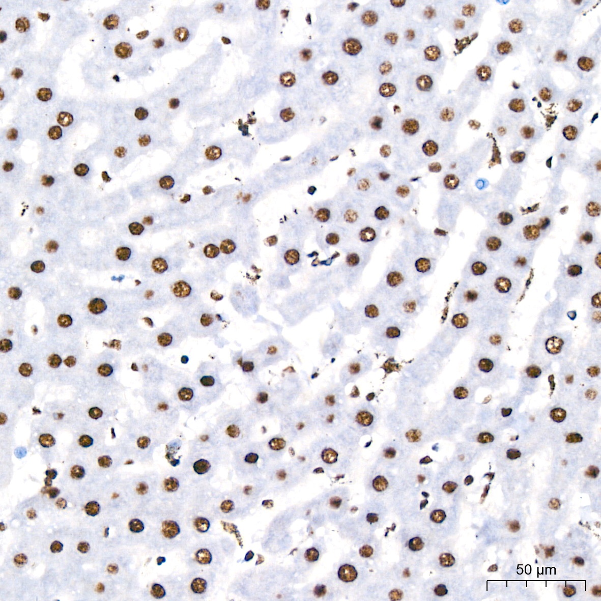
Immunohistochemistry analysis of paraffin-embedded Rat liver tissue using PABPN1 Rabbit mAb (CAB1735) at a dilution of 1:200 (40x lens). High pressure antigen retrieval performed with 0.01M Citrate buffer (pH 6.0) prior to IHC staining.
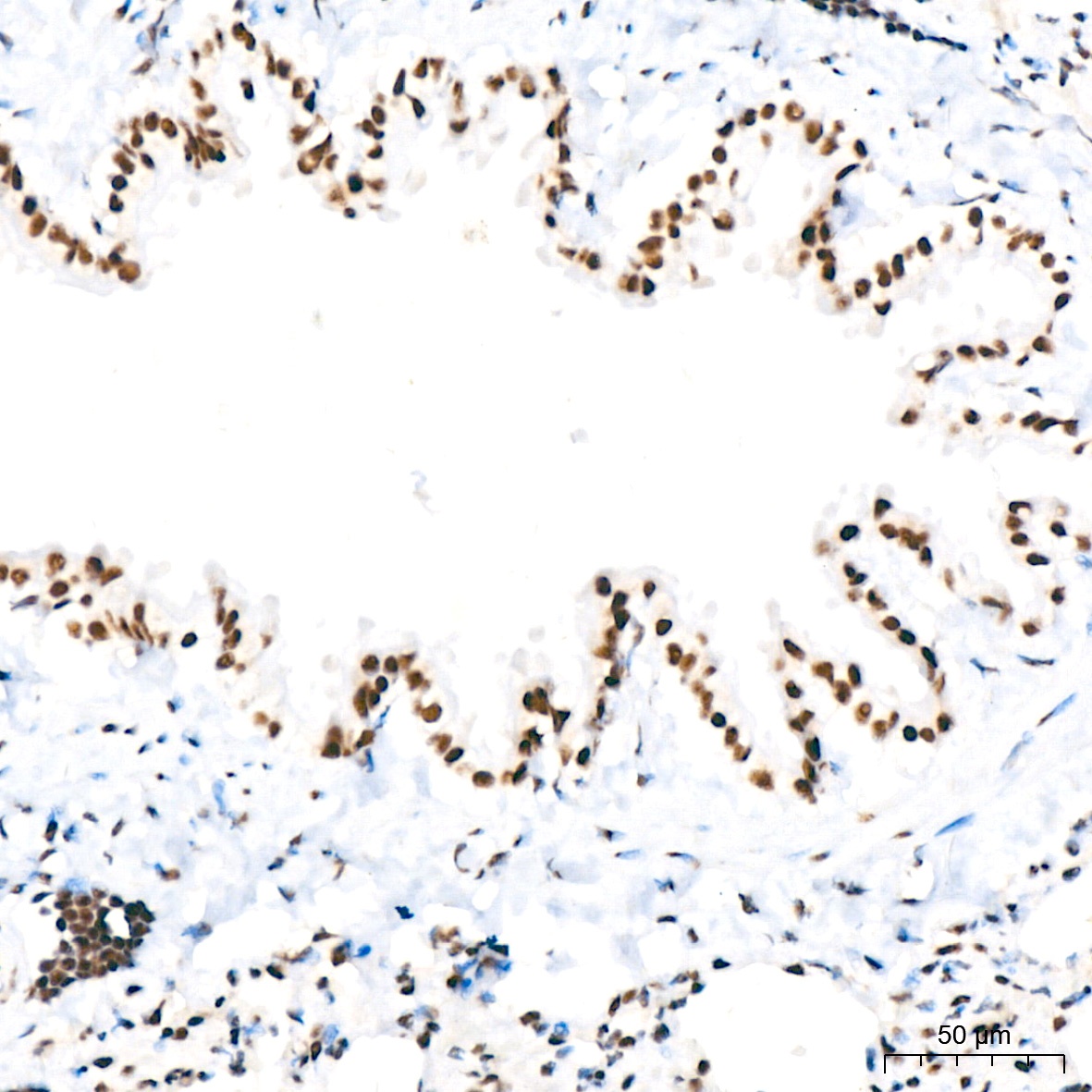
Immunohistochemistry analysis of paraffin-embedded Rat lung tissue using PABPN1 Rabbit mAb (CAB1735) at a dilution of 1:200 (40x lens). High pressure antigen retrieval performed with 0.01M Citrate buffer (pH 6.0) prior to IHC staining.
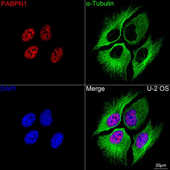
Confocal imaging of U-2 OS cells using PABPN1 Rabbit mAb (CAB1735,dilution 1:200) followed by a further incubation with Cy3 Goat Anti-Rabbit IgG (H+L) (CABS007,dilution 1:500)(Red).The cells were counterstained with α-Tubulin Mouse mAb (AC012, dilution 1:400) followed by incubation with ABflo® 488-conjugated Goat Anti-Mouse IgG (H+L) Ab (CABS076, dilution 1:500) (Green).DAPI was used for nuclear staining (Blue). Objective: 100x.
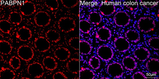
Confocal imaging of paraffin-embedded Human colon cancer using PABPN1 Rabbit mAb (CAB1735, dilution 1:200) followed by a further incubation with Cy3 Goat Anti-Rabbit IgG (H+L) (CABS007, dilution 1:500) (Red). DAPI was used for nuclear staining (Blue). Objective: 40x. Perform high pressure antigen retrieval with 0.01 M citrate buffer (pH 6.0) prior to IF staining.
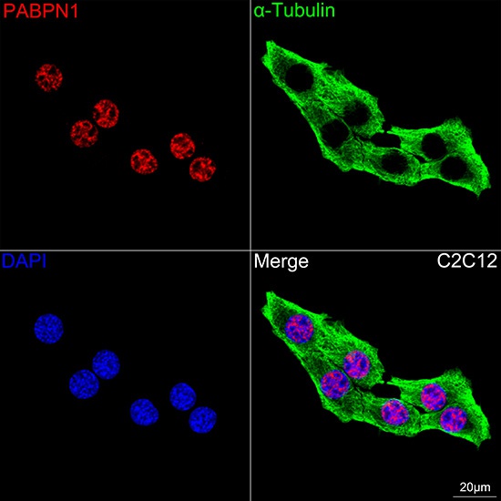
Confocal imaging of C2C12 cells using PABPN1 Rabbit mAb (CAB1735,dilution 1:200) followed by a further incubation with Cy3 Goat Anti-Rabbit IgG (H+L) (CABS007,dilution 1:500)(Red).The cells were counterstained with α-Tubulin Mouse mAb (AC012, dilution 1:400) followed by incubation with ABflo® 488-conjugated Goat Anti-Mouse IgG (H+L) Ab (CABS076, dilution 1:500) (Green).DAPI was used for nuclear staining (Blue). Objective: 100x.


![Anti-PABPN1 [R02-8B1] Monoclonal Antibody (AGMB00578)](https://cdn11.bigcommerce.com/s-h68l9z2lnx/images/stencil/590x590/products/271867/694095/anti-pabpn1-r02-8b1-monoclonal-antibody-agmb00578__75255.1774511372.jpg?c=2 "Anti-PABPN1 [R02-8B1] Monoclonal Antibody (AGMB00578)")


