
Blog
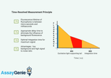
Time-Resolved Fluorescence (TRF): A Comprehensive Introduction
Time-Resolved Fluorescence (TRF) technology is a cutting-edge analytical technique that has revolutionized the fields of biochemistry, immunology, and molecular biology. By offering a refined approach to measuring fluorescent signals, TRF circumvents many of the limitations associated with traditional fluorescence measurements, providing researchers with a tool of unparalleled sensitivity and specificity. Understanding Time-Resolved Fluorescence TRF is based on the principle of delayed fluorescence detection. Unlike conventional fluorescence methods that measure signals immediately after excitation, TRF allows a short delay before measuring the fluorescence. This delay is crucial
…
14th Mar 2024

Understanding Blotchy Western Blots: Causes and Remedies
Western blotting is a powerful and widely used technique in molecular biology, essential for detecting and quantifying specific proteins in complex biological samples. However, despite its popularity, Western blotting can sometimes yield frustrating results, with one of the most common issues being blotchy or uneven bands. Blotchy Western blots can arise from various factors, ranging from sample preparation to technical errors during the blotting process. In this article, we'll delve into the potential causes of blotchy Western blots and provide some tips to rectify them. What are Blotchy Western Blots? Blotchy Western blots refer to the uneven distribution of bands across
…
13th Mar 2024
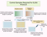
Control Samples Required for ELISA Protocol
Enzyme-Linked Immunosorbent Assay (ELISA) is a powerful analytical biochemistry assay that utilizes antibodies and color change to identify a substance. It is widely used in the field of immunology to detect the presence of antibodies or antigens in a sample. The accuracy and reliability of ELISA depend significantly on the use of appropriate control samples throughout the protocol. This article delves into the essential control samples required for ELISA to ensure the validity and reproducibility of the assay's results. Positive and Negative Controls Positive ControlA positive control is an integral component of the ELISA protocol, serving as a benchmark to confirm that the ass
…
2nd Mar 2024
Fluorescence Resonance Energy Transfer (FRET) Assays: An Insight into Molecular Interactions
Methods · FluorescenceFRET Assays: Principle, Förster Radius and Measurement MethodsFRET transfers energy from an excited donor fluorophore to a nearby acceptor without emitting a photon, and the efficiency falls with the sixth power of the distance between them. That steep dependence is what turns a fluorescence measurement into a molecular ruler with 1–10 nm resolution. This guide covers the physics, how to choose a pair, the four ways FRET is measured, and the controls that separate real signal from bleed-through.Browse fluorescence assays →1–10 nmUsable distance ranger−6How efficiency falls with distanceR0Distance giving 50% transferκ2 = 2/3The usual orientation assumptionKey takeawaysFR
…
1st Mar 2024
Unveiling the Potential of Cy3 Wavelength: Illuminating the Path to Advanced Biomedical Imaging
In the realm of biomedical imaging, the quest for precision and clarity drives researchers to explore innovative technologies. One such breakthrough is the Cy3 wavelength, a powerful tool revolutionizing fluorescence imaging techniques. This article delves into the significance, applications, and advancements of Cy3 wavelength in biomedical research. Understanding Cy3 Wavelength:: Cy3, short for Cyanine 3, is a fluorescent dye belonging to the cyanine dye family. Its exceptional properties make it a popular choice for labeling biomolecules and tracking biological processes under a microscope. Cy3 emits light in the red-orange spectrum, typically around 570 to 590 nanometer
…
23rd Feb 2024
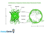
Choosing a Fluorescent Protein: A Comprehensive Guide
Fluorescent proteins (FPs) have revolutionized the field of molecular and cellular biology, allowing scientists to visualize and track biological processes in live cells with unprecedented clarity and specificity. Originating from the green fluorescent protein (GFP) discovered in the jellyfish Aequorea victoria, the palette of available FPs has expanded to include a rainbow of colors. This article provides a detailed overview of factors to consider when choosing a fluorescent protein for your research, ensuring optimal results for your specific applications. Understanding Fluorescent Proteins The Basics of FluorescenceFluorescence occurs when a substance absorbs light at one wav
…
17th Feb 2024
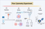
Navigating the Intricacies of Intracellular Flow Cytometry: Key Considerations
In the evolving landscape of cellular biology, intracellular flow cytometry (ICFC) stands out as a powerful technique for analyzing the complexities within cells. This advanced method extends the capabilities of traditional flow cytometry by allowing researchers to investigate intracellular proteins, signaling pathways, and specific gene expressions within individual cells. ICFC has become indispensable in various fields, including immunology, oncology, and drug discovery, offering insights that are pivotal for understanding disease mechanisms and developing therapeutic strategies. However, harnessing the full potential of ICFC requires meticulous attention to several critical considerat
…
16th Feb 2024
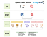
Optimizing Organoid Culture Conditions: Paving the Way for Revolutionary Advances in Biomedical Research
In the dynamic landscape of biomedical research, organoids have emerged as a groundbreaking tool, offering three-dimensional (3D) models that mimic the complex architecture and functionality of human organs. These miniature, self-organizing structures have revolutionized our approach to understanding human development, disease modeling, and drug discovery. However, the key to harnessing their full potential lies in optimizing organoid culture conditions. This intricate process involves fine-tuning the biochemical and physical environment to support the growth, differentiation, and maturation of organoids. This article delves into the critical aspects of organoid culture, including the se
…
14th Feb 2024
What is the difference between PBS and dPBS?
Lab Buffers · ProtocolsPBS vs DPBS: What’s the Difference?PBS and DPBS are the everyday isotonic, pH-balanced salt solutions used to wash, dilute and transport cells and proteins. This guide breaks down their composition, the calcium/magnesium question, and how to choose the right one for your experiment.Browse ELISA Kits & Reagents →7.4PHYSIOLOGICAL pH~300mOsm/kg (ISOTONIC)±Ca/MgDPBS VARIANTS3+COMMON LAB USESQuick answerPBS (phosphate-buffered saline) and DPBS (Dulbecco’s phosphate-buffered saline) are isotonic, pH-7.4 salt solutions used to wash, dilute and transport cells and proteins. The key difference is formulation: standard DPBS is often supplied without calcium and magnesi
…
8th Feb 2024
Transduction vs Transfection: Understanding Gene Delivery Techniques
Techniques · Gene DeliveryTransduction vs Transfection: Understanding Gene Delivery TechniquesTransduction and transfection are the two main routes for introducing genetic material into cells. Transduction uses viral vectors to deliver genes efficiently, even into primary and non-dividing cells, while transfection relies on non-viral chemical or physical methods. This guide compares how they work, their advantages and limitations, and how to choose the right approach.Browse Molecular Biology Tools →ViralTRANSDUCTIONNon-viralTRANSFECTIONStable / transientEXPRESSIONEfficiencyKEY TRADE-OFFContentsTransductionTransfectionApplications in Research and TherapyTransduction vs Transfection at a Glanc
…
8th Feb 2024
Guide to Western Blot Sample Preparation
Protein Analysis · ProtocolsWestern Blot Sample Preparation: A Practical GuideMost failed Western blots fail before the gel is loaded. Lysis buffer choice, inhibitor selection, accurate protein quantification and correct denaturation determine whether a band appears at all — and several of the conventional rules, including boiling every sample at 95 °C, are wrong for particular protein classes. This guide covers each step with the decisions that actually change the outcome.Browse lysis buffers →20–30 µgTypical protein load per lane−80 °CLong-term sample storage95 °CStandard denaturation temperature37–70 °CUse instead for membrane proteinsKey takeawaysMatch the lysis buffer to the target
…
6th Feb 2024
Mouse Injection Guide: Intraperitoneal, Subcutaneous, Intramuscular & Intravenous Routes
In Vivo Methods · ProtocolsMouse Injection Guide: Intraperitoneal, Subcutaneous, Intramuscular & Intravenous RoutesRoute choice and injection technique determine how much of a dose actually reaches its target, how fast, and how much stress the animal experiences. This guide covers route selection, volume and needle-gauge limits, restraint and technique, aftercare, and the readouts used to measure the response.Browse mouse assay kits →10 mL/kgTypical intraperitoneal volume ceiling5 mL/kgTypical intravenous bolus ceiling≤0.05 mLMaximum intramuscular volume per site27–30 GGauge range for tail-vein injectionKey takeawaysRoute determines absorption rate and maximum volume: intraperitoneal and
…
4th Feb 2024
Serum vs Plasma: Key Differences & When to Use Each [Guide]
Sample Preparation · ImmunoassaySerum vs Plasma: Key Differences and When to Use EachSerum and plasma come from the same blood draw but are not interchangeable. One is collected after clotting, the other before it, and that single difference changes which analytes survive, which are artefactually raised, and which assays will give you a trustworthy number. This guide covers the composition difference, collection protocols for both, tube selection, and how to choose the right matrix for an ELISA.Browse ELISA kits →15–30 minClotting time before serum can be spun≤2 hWindow to separate plasma from cells1,000–2,000 ×gTypical centrifugation for separation~15–20%Greater yield from plasma than serum
…
24th Jan 2024
Hybridoma Technology: Revolutionizing Antibody Production
Immunology · Antibody ProductionHybridoma Technology: How Monoclonal Antibodies Are MadeHybridoma technology, pioneered by Georges Köhler and César Milstein in 1975, is the method that made monoclonal antibodies possible. By fusing an antibody-producing B cell with an immortal myeloma cell, it creates a hybrid that churns out a single, defined antibody indefinitely — the foundation of modern diagnostics, therapeutics and research.Explore the antibody range →1975Köhler & Milstein invent the method1984Nobel Prize awarded1antibody per hybridoma clone∞immortal, continuous productionKey TakeawaysHybridoma technology fuses a B cell with a myeloma cell to make an immortal, antibody-producing hy
…
23rd Jan 2024
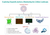
Exploring Organelle Markers: Illuminating the Cellular Landscape
The eukaryotic cell is a complex and dynamic system with distinct membrane-bound compartments known as organelles, each performing specialized functions essential for cellular survival. To better understand the intricate workings of these organelles, scientists employ organelle markers – molecular tools that provide a means to visualize and study these structures. In this article, we delve into the fascinating realm of organelle markers, discussing their significance, types, and applications in unraveling the mysteries of cellular biology. Organelle Markers: Painting a Molecular Canvas Organelle markers play a pivotal role in cell biology research by allowing scientists to
…
11th Jan 2024

In Vitro vs In Vivo: Complete Comparison + Selection Guide | Research Methods
Discover the distinct roles of 'In Vivo' and 'In Vitro' in biological research, where one offers a natural context and the other precise control, both vital for scientific advancement. Key Takeaways: "In Vivo" studies are within living organisms, offering physiological relevance. "In Vitro" studies occur outside organisms, allowing controlled experiments. Both methods are complementary, each providing unique scientific insights. "In Situ" studies observe phenomena in their natural environments. In the field of biological research, scientists employ various experimental methods to understand complex biological processes. Two fundamental approaches at the forefron
…
2nd Aug 2023

Transfection: A Comprehensive Guide
Transfection protocols play a crucial role in the realm of molecular biology and genetic research, enabling the introduction of foreign nucleic acids into living cells. This innovative technique allows scientists to manipulate gene expression, study protein function, and investigate cellular processes with remarkable precision. This article focusses on the fundamental principles underlying transfection protocols, exploring their significance and various approaches employed for achieving successful genetic delivery
Table of Contents
Jump to a section:
- Cell Transfection
- Applications
- Mechanism
- Transfection vs Tr
…
12th Jul 2023

Transformation Protocol – Sample Transformation Protocols
Introduction Transformation of bacteria with plasmids is not only of note for bacterial studies but also can be used for gene expression studies in mammalian cells. Most plasmids are of bacterial origin and contain both a bacterial origin of replication and an antibiotic resistance gene which can be used as a selectable marker. The process of transformation allows foreign DNA to be introduced into a cell. Genetic modifications can be made to various bacterial strains to make them more amenable to transformation. Such modifications will maintain the plasmid without rearranging the plasmid DNA. Certain treatments have been shown to increase the transformation efficiency of bacteria.
…
27th Jun 2023

Mycoplasma in Cell Culture Detection Guide
As a PhD student delving deep into the realm of cellular biology, there is one antagonist that consistently challenges my research: mycoplasma. This microscopic bacterium, with its ability to invisibly infiltrate and undermine cell cultures, has become a persistent foe. However, understanding the intricacies of this bacterial contaminant is the first step towards effective mitigation.
window.SHOGUN_IMAGE_V2_ELEMENTS = window.SHOGUN_IMAGE_V2_ELEMENTS || new Array();
window.SHOGUN_IMAGE_V2_ELEMENTS.push({ hoverImage: '', uuid: 's-9662d6e3-0a1e-4340-82be-21f2d6baf7d7' })
Schematic of Mycoplasma
The Invisible Enemy: Understan
…
28th May 2023
Renal and Hepatic Function Assays
Hepatic Function Assays
Liver function assays are a suite of tests designed to assess the health and efficacy of our vital organ: The liver. This hardworking powerhouse is responsible for creating bile, eliminating toxins, and breaking down fats, carbohydrates and proteins – all necessary elements that keep us ticking!
To evaluate the liver's health, there are various hepatic function assays that can be performed. These tests measure the organ's capacity to produce bile, metabolize toxins and drugs, as well as synthesize proteins and carbohydrates. By assessing these three factors of liver performance with an
…
7th Mar 2023
Flow Cytometry Protocol | 10 Hints & Tips
Lab Methods · Flow CytometryFlow Cytometry Protocol: Two Worked Protocols, Controls and 10 Practical TipsFlow cytometry measures individual cells in a fluid stream at hundreds per second, reporting size, granularity and fluorescence for each one. Most failures come not from the instrument but from sample preparation, panel design and controls. This guide gives two complete worked protocols, a full controls reference table, and ten practical points that determine whether the data is interpretable.Browse flow cytometry reagents →FSC / SSCSize and granularityPMTPhotomultiplier voltageFMOThe control that sets gates9Controls in the reference tableKey takeawaysForward scatter correlates with parti
…
7th Nov 2022
Cell Synchronisation Methods
Cell Culture · ProtocolsCell Synchronisation Methods: Protocols for G1, S, G2 and M Phase ArrestAn asynchronous culture contains cells at every stage of the cycle at once, so any measurement is an average across phases. Synchronisation forces cells into one phase so that phase-specific events can actually be observed. This guide covers the chemical and physical approaches, six working protocols with concentrations and timings, how to confirm synchrony by flow cytometry, and the artefacts these methods introduce.Browse cell cycle reagents →2 mMThymidine, double block100 nMNocodazole for M-phase arrest9 µMRO-3306 for G2/M arrestFlow + PIStandard synchrony confirmationKey takeawaysSynchronisati
…
26th Oct 2022
Guide to Fluorescence Activated Cell Sorting (FACs): The Latest Technology in Cell Separation
Flow Cytometry · Cell SortingGuide to Fluorescence-Activated Cell Sorting (FACS)Fluorescence-activated cell sorting (FACS) is a specialised form of flow cytometry that physically isolates individual cells from a mixed sample based on their fluorescence and light-scattering properties. With sorting purities approaching 99.99%, it is one of the most powerful tools in modern cell biology, immunology and stem cell research.Explore flow cytometry antibodies →99.99%achievable sort purity1000sof cells sorted per second4+parameters measured at onceSinglecell resolutionKey TakeawaysFACS isolates individual cells with a purity approaching 99.99%.It sorts cells by size, granularity, surface markers and
…
26th Jul 2022

RIPA Buffer Recipe - Cell Lysis | Assay Genie
RIPA (Radio Immuno Precipitation Assay) buffer is mostly used when carrying out a western blot or immunoprecipatation assay. A RIPA buffer is used in order to lyse cells and extract protein from cultured cells. RIPA buffer cell lysis enables determination of protein concentration. RIPA buffer is an ideal cell lysis reagent since it contains three non-ionic and ionic detergents.
Preserving protein:protein interactions
When you need to preserve protein-protein interactions or to reduce denaturation its recommended to use a RIPA buffer recipe without SDS (ionic detergent) or Triton X-100 (non-ionic detergent).
RIPA buffer recipes
…
20th Aug 2021