

Fatty Acid Oxidation Assay Kit: 20 Published Applications

Summary
The Assay Genie Fatty Acid Oxidation (FAO) Assay Kit (BR00001) is a colorimetric plate-reader assay. Cell or tissue lysate oxidises an octanoyl-CoA substrate, and the NADH produced reduces the tetrazolium dye INT to a red formazan, read at 492 nm. The 20 studies summarised here used it on primary hepatocytes, myotubes, cardiomyocytes, podocytes, immune cells, neuronal cells, isolated adipocyte mitochondria and tissue lysates (muscle, adipose, kidney cortex, placenta, fish liver). In most of them, the FAO readout was the functional check on a gene- or protein-level change in lipid metabolism.
Key takeaways
- 20 published studies used BR00001 across liver, skeletal muscle, heart, adipose tissue, kidney, immune cells, neuronal cells, placenta and farmed fish.
- Samples ranged from intact cultured cells to frozen tissue lysates and isolated mitochondrial fractions.
- Most groups normalised to total protein (BCA) and paired the FAO readout with CPT1/PPARα manipulation, oxygen consumption or gene expression.
- The homogenate format measures β-oxidation enzyme capacity, not CPT1-gated flux in intact cells; studies linking FAO to CPT1 relied on knockdown or inhibitor data.
Product cited in these studies
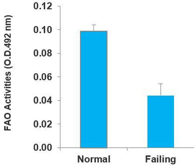
Fatty Acid Oxidation (FAO) Assay Kit (BR00001)
Used in the 20 studies below to measure β-oxidation activity in cell and tissue lysates.
View kit →At a glance
| Study | Model / species | Sample type | Product | What it measured | Role in study |
|---|---|---|---|---|---|
| Papa et al., 2026, Cell Metab [1] | Primary mouse hepatocytes; bempedoic acid ± Ppara siRNA | Hepatocytes in 6-well plates | BR00001 | FAO activity, protein-normalised | Showed drug-induced FAO needs PPARα |
| Wang J et al., 2026, J Pharm Pharmacol [2] | MCD-diet MASH model (species not in abstract) | Not accessible | BR00001 (per citation record) | FAO activity | FAO restored by CGA treatment |
| Zhu et al., 2025, Histol Histopathol [3] | Male C57BL/6 mice, 12-week HFD MASLD | Not accessible | Assay Genie FAO kit | FAO rate | Part of PPARα/CPT1α mechanism work |
| Wang Y et al., 2024, Free Radic Biol Med [4] | Terf2ip KO vs WT mice, HFD or MCD | Not accessible | Assay Genie FAO kit | FAO | FAO impaired by Terf2ip loss |
| Odria et al., 2025, GeroScience [5] | Old (20–22 mo) WT vs HDAC11−/− mice | Protein extract of frozen skeletal muscle | BR00001 | FAO (ΔOD) | Enhanced muscle FAO without HDAC11 |
| Park et al., 2025, J Cachexia Sarcopenia Muscle [6] | C2C12 and human skeletal muscle (HSKMC) myotubes | Cell lysates | BR00001 | FAO activity | Celecoxib raised myotube FAO |
| Odera et al., 2026, Food Funct [7] | SAMP8 mice fed PQQ or IPQ | Not accessible (citation record lists liver) | Assay Genie FAO kit, octanoyl-CoA | FAO | Part of ageing phenotype panel |
| She et al., 2025, Nat Commun [8] | Neonatal rat ventricular myocytes (NRVMs), Hey2 overexpression | Lysate of 1 × 10⁵ NRVMs | BR00001 | FAO enzyme activity | FAO fell with HEY2 induction |
| Chen et al., 2025, Theranostics [9] | Neonatal mouse cardiomyocytes, Ang II, CPT1b knockdown | Cell lysates | BR00001 | FAO, units/µg protein | CPT1b knockdown lowered FAO |
| Natarajan et al., 2024, Aging Cell [10] | 18-month-old mice, chronic β3-AR agonist CL316,243 | Lysates of frozen BAT, iWAT, eWAT | Assay Genie FAO kit | FAO, units/µg protein | FAO rose in iWAT, not BAT |
| Fuwa et al., 2024, FEBS Lett [11] | Male C57BL/6 mice, epididymal white adipocytes | Isolated mitochondrial fractions (CMw, PDMw) | Assay Genie FAO kit | β-oxidation activity | Cytoplasmic > peridroplet mitochondria |
| Wu et al., 2024, Diabetologia [12] | Mouse podocytes ± PCSK9 over/under-expression | Lysate of 2 × 10⁵ podocytes | BR00001 | FAO | Low PCSK9 worsened FAO loss |
| Standage et al., 2021, Am J Physiol Renal [13] | Male C57BL/6J mice, CLP sepsis | Renal cortex | Assay Genie FAO kit | Renal cortical FAO | Low FAO tracked with creatinine |
| Fu et al., 2025, Mol Med [14] | PMN-MDSCs from 4-NQO oesophageal cancer mice | Sorted splenic PMN-MDSCs | BR00001 | FAO level | PKN2 raised CPT1B-dependent FAO |
| Huang et al., 2026, Biomater Res [15] | RAW264.7 macrophages ± LGG nanovesicles | Cells in 96-well plates | BR00001 | Cellular FAO | Vesicles raised FAO via AhR/NQO1 |
| Lei et al., 2026, Commun Biol [16] | C. trachomatis-infected host cells (details not accessible) | Not accessible | Assay Genie FAO kit | FAO | Part of lipid-scavenging mechanism |
| Quentin et al., 2024, J Mitochondria Plastids Endosymbiosis [17] | Human SH-SY5Y-APP695 vs MOCK cells, OEA | Cells in 96-well plates | BR00001 | FAO | OEA raised FAO in APP695 cells only |
| Toledano et al., 2026, Research Square preprint [18] | Human placentas, advanced maternal age vs controls | Placental tissue | BR00001 | FAO activity | Higher FAO with advanced maternal age |
| Kim S, Lim H et al., 2026, Aquac Nutr [19] | Atlantic salmon, fish-oil replacement at 24–25 °C | Liver homogenate | BR00001 | Hepatic β-oxidation | Rose with oil replacement |
| Kim S, Lim J et al., 2026, Mar Biotechnol [20] | Juvenile olive flounder, dietary pantothenic acid | Liver | BR00001 | Hepatic β-oxidation | Higher with pantothenic acid |
Why measure fatty acid oxidation?
Mitochondrial β-oxidation breaks fatty acids into acetyl-CoA, producing NADH and FADH₂ for the electron transport chain. Long-chain fatty acids are first activated to acyl-CoA. CPT1A (liver and most tissues) or CPT1B (muscle and heart) then converts them to acylcarnitines, which cross the inner membrane and are reconverted to acyl-CoA by CPT2. Each turn of the spiral runs four steps: dehydrogenation by acyl-CoA dehydrogenases such as ACADM, hydration, a second dehydrogenation and thiolysis. For long-chain substrates, the last three steps are catalysed by the mitochondrial trifunctional protein, whose α-subunit is HADHA.
Transcriptional control runs largely through PPARα and the coactivator PGC-1α (PPARGC1A). Many papers infer FAO from mRNA for these genes, but transcript levels do not prove pathway activity. An enzyme-activity readout lets researchers test whether a change in expression changes oxidative capacity. That pattern recurs in the studies below.

How the assay works
Per the product page, each lysate goes into two wells: one with octanoyl-CoA reaction solution and one with substrate-free control solution. After 1–2 h at 37 °C the reaction is stopped with 3% acetic acid and read at 492 nm. Subtracting the control well removes NADH from endogenous substrates; ΔOD is converted to enzyme units (there is no standard curve). The page also notes that CPT activity cannot be measured in this homogenate format, so the readout reflects β-oxidation enzyme capacity, not CPT1-gated flux in intact cells.

How the product was used
Liver, MASLD and hepatic lipid metabolism
Papa et al. (2026) asked how bempedoic acid lowers hepatic lipids. Primary mouse hepatocytes (10⁶ per well) received Ppara or control siRNA, then bempedoic acid, the PPARα agonist GW7647 or vehicle for 48 h, followed by a 60-min kit reaction; ΔOD-derived activity was normalised to BCA protein. Bempedoic acid raised FAO to levels comparable with GW7647, and Ppara knockdown abolished the rise, the functional evidence for PPARα dependence [1].
Wang J et al. (2026) tested a three-component herbal combination (CGA) in a methionine–choline-deficient MASH model. The abstract reports that CGA restored FAO alongside PPARα and FXR. The authors placed FAO recovery downstream of FXR–PPARα activation. Methods detail not accessible [2].
Zhu et al. (2025) studied licochalcone A in male C57BL/6 mice fed a high-fat diet for 12 weeks. A PPARα inhibitor abolished its protective effect. The FAO rate was assessed with the Assay Genie kit, but the sample type was not accessible [3].
Wang Y et al. (2024) fed Terf2ip knockout and wild-type mice a high-fat or MCD diet. They reported that loss of TERF2IP impaired lipophagy and FAO through SIRT1/AMPK signalling. Methods detail not accessible [4].
Skeletal muscle and ageing
Odria et al. (2025) compared old (20–22 months) wild-type and HDAC11-deficient mice. FAO was measured in protein extracts of flash-frozen skeletal muscle, with control readings subtracted from reaction readings. FAO activity was significantly higher without HDAC11. Fewer lipid droplets in soleus, higher Cpt1b, Acot1 and Ucp3 expression and smaller age-related shifts in muscle fatty acid composition supported the result [5].
Park et al. (2025) screened for agonists of the prokineticin receptor PROKR1 and identified celecoxib. Lysates of C2C12 and human skeletal muscle myotubes were incubated with 50 µl reaction solution at 37 °C for 2 h. Celecoxib increased FAO activity in both, alongside higher mitochondrial content, supporting the in vivo finding of more oxidative fibres in offspring of treated dams [6].
Odera et al. (2026) fed senescence-accelerated SAMP8 mice pyrroloquinoline quinone (PQQ) or imidazopyrroloquinoline (IPQ) over their lifetime or from midlife. The citation snippet states that FAO was measured with the Assay Genie kit using octanoyl-CoA as substrate, and the chemRxiv preprint of the same study [7b] carries the same statement. The abstract reports better muscle function with both compounds and less lipid in adipose tissue and liver with IPQ. Methods detail not accessible [7].
Heart
She et al. (2025) asked how the transcriptional repressor HEY2 affects cardiomyocyte energetics. Lysates of 1 × 10⁵ neonatal rat ventricular myocytes were read at 492 nm. FAO activity fell with adenoviral Hey2 overexpression, matching lower PGC-1α, ESRRA and CPT1 protein and reduced mitochondrial mass [8].
Chen et al. (2025) studied the circular RNA CHACR in cardiac hypertrophy (TAC mice; angiotensin II-treated neonatal mouse cardiomyocytes). Lysate (20 µl) was run in duplicate for 60 min at 37 °C and expressed as units/µg protein. CPT1b knockdown reduced FAO and ATP (supplementary data); FAO was not measured with CHACR manipulation, and the final mechanism ran through L-carnitine and Jak2/Stat3 [9].
Adipose tissue and thermogenesis
Natarajan et al. (2024) gave 18-month-old mice the β3-adrenergic agonist CL316,243 by osmotic minipump for 4 weeks. FAO was measured in lysates of frozen adipose tissue and expressed as units/µg protein. It increased in inguinal WAT but, unexpectedly, not in BAT. Epididymal WAT fell below the assay's detection limit. Together with UCP1 data and lipid-turnover tracing, this supported a UCP1-independent route to energy expenditure in white fat [10].
Fuwa et al. (2024) separated cytoplasmic (CMw) and peridroplet (PDMw) mitochondria from epididymal white adipocytes by centrifugation. β-oxidation activity, measured with the kit, was higher in CMw, while citrate synthase activity and cytochrome c content were similar. Higher HADH and ACAA1 in CMw agreed. PDMw were associated with diacylglycerol acyltransferase 2, and the authors proposed roles for CMw in β-oxidation and for PDMw in lipid-droplet expansion [11].
Kidney
Wu et al. (2024) studied PCSK9 in diabetic kidney disease. Lysates of 2 × 10⁵ podocytes were protein-assayed by BCA and run in duplicate (control and reaction wells). Plates were read at 30 and 60 min. TNF-α or palmitic acid reduced FAO; Pcsk9 siRNA made it lowest and PCSK9 overexpression partly restored it, in line with Ppargc1a, Cpt1 and Acox1 expression [12].
Standage et al. (2021) combined serum and urine NMR metabolomics with renal cortical FAO in mice 24 h after caecal ligation and puncture. FAO did not differ between septic and sham mice overall. In septic mice, however, FAO fell as serum cytokines rose, and lower FAO went with higher serum creatinine. In septic mice, FAO also correlated positively with FAO-gene expression [13].
Immune cells
Fu et al. (2025) asked how PKN2 boosts the immunosuppressive activity of polymorphonuclear myeloid-derived suppressor cells in oesophageal cancer. PMN-MDSCs were magnetically sorted from spleens of 4-NQO-treated mice and given a PKN2 lentivirus. PKN2 raised FAO in the kit assay and FAO-dependent oxygen consumption (Seahorse, with etomoxir). CPT1B siRNA reduced the PKN2-induced rise in both readouts, whereas CPT1A or CPT1C siRNA did not change oxygen consumption [14].
Huang et al. (2026) treated RAW264.7 macrophages, with or without LPS, with Lactobacillus rhamnosus GG-derived nanovesicles. Vesicles increased FAO under control and inflammatory conditions. An AhR inhibitor or NQO1 knockdown reduced this, while NADH enhanced it, supporting an AhR/NQO1/CPT1A axis behind reparative macrophage polarisation and bone regeneration in periodontitis [15].
Infection, neuronal and placental models
Lei et al. (2026) reported that Chlamydia trachomatis induces lipophagy through METTL14-mediated m⁶A modification of PPARα mRNA to support its replication. The citation record shows an Assay Genie FAO kit was used. The host-cell model and methods detail were not accessible [16].
Quentin et al. (2024) treated SH-SY5Y-APP695 cells (an early Alzheimer's model) and vector controls with oleoylethanolamide. Cells were seeded at 2 × 10⁴ per well and the kit reaction ran for 90 min. Two doses of 0.05 µM raised FAO by 18.4% in APP695 cells, with no change in controls. CPT1 mRNA was unchanged. With higher ATP and lactate/pyruvate ratio, the authors linked the extra ATP in APP695 cells to FAO and glycolysis [17].
Toledano et al. (2026, preprint) compared placentas from uncomplicated pregnancies with and without advanced maternal age. FAO activity and PPAR-γ expression were higher in the older-mother group, despite lower PGC-1α, NRF2 and TFAM. The authors interpreted this as a shift towards lipid-based energy metabolism. Methods detail not accessible [18].
Aquaculture nutrition
Kim S, Lim H et al. (2026) replaced fish oil with a Schizochytrium–canola oil blend (0–100%) in Atlantic salmon held at 24–25 °C for 6 weeks. Liver (20 mg) was homogenised, adjusted to 1–2 mg/ml protein and run in duplicate for 60–120 min. β-oxidation was significantly higher at 40–100% replacement, matching higher ACOX1, CPT1 and PPARα expression [19].
Kim S, Lim J et al. (2026) fed juvenile olive flounder 0–60 mg/kg pantothenic acid, the CoA precursor, for 12 weeks. Hepatic β-oxidation activity, CoA and pantothenic acid were higher in all supplemented groups than in the basal-diet group, while liver lipid was lower. Methods detail not accessible beyond the kit statement [20].
Cross-study insights
- Sample types. Intact cultured cells (hepatocytes, myotubes, cardiomyocytes, podocytes, MDSCs, macrophages, neuronal cells), frozen tissue lysates (muscle, adipose, renal cortex, placenta, fish liver) and isolated mitochondrial fractions [11] all gave usable readouts. Natarajan et al. report the one clear limitation: epididymal WAT fell below the detection limit [10].
- Normalisation. Most groups measured total protein by BCA and reported activity per µg protein [1, 9, 10] or adjusted protein input [19]. Others fixed cell number [8, 17]. Fuwa et al. measured cytochrome c in parallel, separating mitochondrial content from activity per mitochondrion [11].
- Incubation time. Reported times ranged from 30–60 min [12] to 2 h [6], within the product's 1–2 h range. Reading at more than one time point [12] helps confirm the signal is still linear.
- Pairing with other readouts. The assay was rarely used alone. Partners included CPT1/PPARα genetic manipulation [1, 9, 14], etomoxir-sensitive OCR [14], citrate synthase [11, 18], ATP [9, 17] and FAO-gene expression [5, 12, 19].
- Null results. No overall sham–sepsis difference [13], no BAT increase [10] and no change in control neuronal cells [17] show that lysate capacity does not always track gene expression or whole-body energy expenditure.
- CPT1 interpretation. Several studies linked FAO changes to CPT1 [9, 14, 15]. Because the homogenate format with octanoyl-CoA does not measure CPT activity, those conclusions rested on CPT1 knockdown or inhibitor data, not on the FAO readout alone.
Practical considerations
Generic best practice and product-page guidance, not reported by the cited studies:
- Prepare samples cold and fast. The product page recommends about 10⁶ cells, or about 25 mg tissue per 0.5 ml lysis solution, protein adjusted to 1–3 mg/ml. Avoid reducing agents and SDS, and avoid repeated freeze–thaw, which can reduce activity.
- Remove blood. Wash tissue well; the page warns that blood cells give inconsistent results and that red colour interferes at 492 nm. Extracellular fluids are not suitable.
- Controls. Run the paired substrate-free well for every sample, and include a high-mitochondria tissue or cell lysate as a positive control. The product page specifies a humidified 37 °C incubator, not a CO₂ incubator.
- Pair with a flux method when it matters. Use Seahorse or radiolabelled palmitate when CPT1-dependent, long-chain oxidation in intact cells is the question.
Measuring fatty acid oxidation?
The colorimetric FAO assay kit used in these 20 studies is available from Assay Genie, with expert technical support.
View the FAO Assay Kit →Frequently asked questions
How do you measure fatty acid oxidation in cells or tissue?
Common options are colorimetric lysate assays, extracellular flux (oxygen consumption with etomoxir), radiolabelled palmitate oxidation and live-cell luminescent assays. A colorimetric kit such as BR00001 measures β-oxidation enzyme capacity in lysates on a standard plate reader. It suits comparing many tissue or cell samples, and frozen tissue can be used.
What does the BR00001 FAO assay actually measure?
It measures how fast enzymes in a lysate oxidise the medium-chain substrate octanoyl-CoA. NADH from the reaction reduces INT to a red formazan read at 492 nm. A control well without substrate is subtracted. Because the cell structure is lost, the assay does not capture CPT1-dependent transport of long-chain fatty acids.
Can I use frozen tissue for a fatty acid oxidation assay?
Yes. The product page states that the assay works with frozen tissue because it measures enzyme activity, and published studies used frozen skeletal muscle and adipose tissue. Keep lysates on ice, avoid repeated freeze–thaw cycles and normalise to total protein so samples processed on different days can be compared.
How should FAO assay results be normalised?
Most published users measured protein by BCA and reported activity per µg protein, or loaded equal protein. Cell number is an alternative for cultured cells. When comparing mitochondrial fractions, a mitochondrial marker such as cytochrome c or citrate synthase activity separates differences in mitochondrial content from differences in activity per mitochondrion.
Is a colorimetric FAO assay the same as a Seahorse assay?
No. Seahorse infers FAO from oxygen consumption in intact, respiring cells, using inhibitors such as etomoxir to show specificity. A colorimetric kit is an endpoint enzyme-capacity assay on lysates. The methods answer different questions, and some studies, such as Fu et al. (2025), used both.
References
- Papa C, Rose A, Martin HNG, et al. Bempedoic acid directly binds and activates PPARα. Cell Metabolism. 2026;38(4):824–830.e5. doi:10.1016/j.cmet.2025.12.018.
- Wang JJ, Yang HL, Zhang DQ, et al. The CGA combination enhances bile acids alternative biosynthesis pathway and alleviates MASH induced by methionine-choline-deficient diet. Journal of Pharmacy and Pharmacology. 2026;78(3):rgaf124. doi:10.1093/jpp/rgaf124.
- Zhu W, Xu H, Yan R, et al. Licochalcone A ameliorates lipid accumulation in metabolic dysfunction-associated steatotic liver disease via upregulating PPARα/CPT1α. Histology and Histopathology. 2025;40(12):2011–2023. doi:10.14670/HH-18-907.
- Wang Y, Liu S, Ni M, et al. Terf2ip deficiency accelerates non-alcoholic steatohepatitis through regulating lipophagy and fatty acid oxidation via Sirt1/AMPK pathway. Free Radical Biology and Medicine. 2024;220:78–91. doi:10.1016/j.freeradbiomed.2024.04.238.
- Odria R, Cardús A, Gomis-Coloma C, et al. HDAC11 deficiency regulates age-related muscle decline and sarcopenia. GeroScience. 2025;47(4):5843–5868. doi:10.1007/s11357-025-01611-y.
- Park JH, Mok J, Park S, et al. Celecoxib enhances oxidative muscle fibre formation and improves muscle functions through Prokr1 activation in mice. Journal of Cachexia, Sarcopenia and Muscle. 2025;16(1):e13704. doi:10.1002/jcsm.13704.
- Odera K, Tanaka Y, Ikemoto K, Tamakoshi M, Takahashi R. Pyrroloquinoline quinone and imidazopyrroloquinoline intake diminish mortality risk during midlife and improve muscular dysfunctions with age in mice. Food & Function. 2026;17(14):6343–6358. doi:10.1039/d6fo00788k. (7b: preprint of the same study, ChemRxiv 2026, doi:10.26434/chemrxiv.15000199/v1.)
- She P, Gao B, Li D, et al. The transcriptional repressor HEY2 regulates mitochondrial oxidative respiration to maintain cardiac homeostasis. Nature Communications. 2025;16:232. doi:10.1038/s41467-024-55557-4.
- Chen L, Wang W, Zhao Y, Zhang S, Zhou X. Circular RNA CHACR is involved in the pathogenesis of cardiac hypertrophy. Theranostics. 2025;15(8):3627–3642. doi:10.7150/thno.104695.
- Natarajan D, Plakkot B, Tiwari K, et al. Chronic β3-AR stimulation activates distinct thermogenic mechanisms in brown and white adipose tissue and improves systemic metabolism in aged mice. Aging Cell. 2024;23(12):e14321. doi:10.1111/acel.14321.
- Fuwa M, Kajita K, Mori I, et al. Mitochondrial fractions located in the cytoplasmic and peridroplet areas of white adipocytes have distinct roles. FEBS Letters. 2024;598(14):1753–1768. doi:10.1002/1873-3468.14877.
- Wu M, Yoon CY, Park J, et al. The role of PCSK9 in glomerular lipid accumulation and renal injury in diabetic kidney disease. Diabetologia. 2024;67:1980–1997. doi:10.1007/s00125-024-06191-8.
- Standage SW, Xu S, Brown L, et al. NMR-based serum and urine metabolomic profile reveals suppression of mitochondrial pathways in experimental sepsis-associated acute kidney injury. American Journal of Physiology – Renal Physiology. 2021;320(5):F984–F1000. doi:10.1152/ajprenal.00582.2020.
- Fu X, Zhang GQ, Hou ZC, Fu TT, Cui GH. PKN2 enhances the immunosuppressive activity of polymorphonuclear myeloid-derived suppressor cells in esophageal carcinoma by mediating fatty acid oxidation. Molecular Medicine. 2025;31(1):92. doi:10.1186/s10020-025-01132-6.
- Huang Z, Liu Y, Yang Q, et al. Tryptophan-enriched Lactobacillus rhamnosus GG-derived nanovesicles promote alveolar bone regeneration through macrophage fatty acid oxidation. Biomaterials Research. 2026;30:0370. doi:10.34133/bmr.0370.
- Lei W, Huang Q, Wen Y, Yang Y, Wu H, Li Z. C. trachomatis modulates host lipid metabolism through METTL14–m6A–PPARα axis for reproduction. Communications Biology. 2026. doi:10.1038/s42003-026-10819-3.
- Quentin A, Dieter F, Reutzel M, Eckert GP. Oleoylethanolamide improves energy disposal in a cellular model of Alzheimer's disease. Journal of Mitochondria, Plastids and Endosymbiosis. 2024;2(1):2317246. doi:10.1080/28347056.2024.2317246.
- Toledano JM, Puche-Juarez M, de Paco Matallana C, et al. Advanced maternal age impairs mitochondrial regulation and energy metabolism in the human placenta. Research Square (preprint, not peer reviewed). 2026. doi:10.21203/rs.3.rs-9611501/v1.
- Kim S, Lim H, Jeon JY, Lee KJ. Combined Schizochytrium and canola oil effectively replaces fish oil in Atlantic salmon (Salmo salar) under chronic thermal stress. Aquaculture Nutrition. 2026;2026:8779572. doi:10.1155/anu/8779572.
- Kim S, Lim J, Lee Y, et al. Determination of dietary pantothenic acid requirement for olive flounder (Paralichthys olivaceus): growth and physiological responses. Marine Biotechnology. 2026;28:70. doi:10.1007/s10126-026-10619-0.
Recent Posts
-
Fatty Acid Oxidation Assay Kit: 20 Published Applications
SummaryThe Assay Genie Fatty Acid Oxidation (FAO) Assay Kit (BR00001) is a colorimetric plate-rea …20th Nov 2026 -
Mycoplasma Detection Kits for Cell Culture: A Guide
SummaryA mycoplasma detection kit screens cell-culture supernatant for mycoplasma DNA so contaminate …20th Nov 2026 -
Kynurenine ELISA in Mouse Microglia, Muscle and Serum
SummaryAssay Genie's Mouse KYN (Kynurenine) ELISA Kit (MOFI01315) has been used to measure kynure …20th Nov 2026