
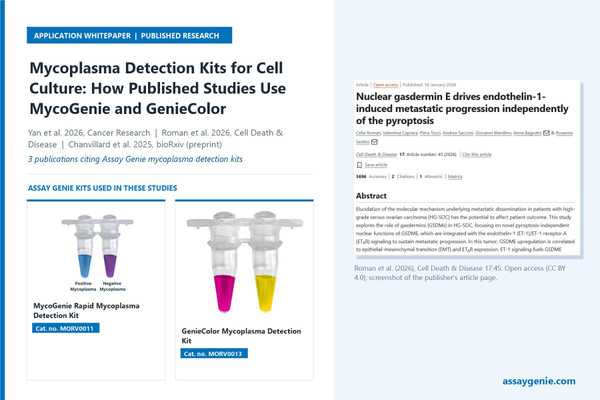
Mycoplasma Detection Kits for Cell Culture: A Guide

Summary
A mycoplasma detection kit screens cell-culture supernatant for mycoplasma DNA so contaminated cultures can be found before they distort experimental data. Assay Genie offers two one-hour, visual isothermal tests: the MycoGenie Rapid Mycoplasma Detection Kit (MORV0011), which detects 28 species and reads purple-to-sky-blue, and the GenieColor Mycoplasma Detection Kit (MORV0013), which detects 40 species and reads pink-to-yellow. Three studies from 2025–26, on liver cancer, ovarian cancer and kidney disease, cite these kits for mycoplasma quality control of their human and mouse cell cultures.
Key takeaways
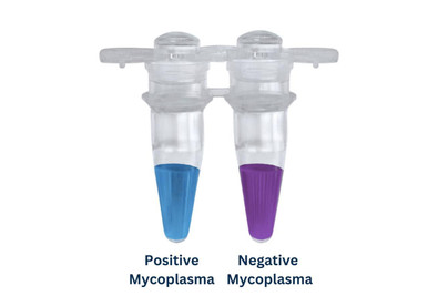
- MycoGenie (MORV0011) detects 28 species and reads purple to sky blue; GenieColor (MORV0013) detects 40 species and reads pink to yellow.
- Both kits amplify mycoplasma DNA from 1 µl of culture supernatant in a single 60-minute incubation, with no thermal cycling, gel or plate reader.
- Three 2025–26 studies on liver cancer, ovarian cancer and kidney disease cite the kits for mycoplasma QC of human and mouse cell cultures.
- Neither paper with readable Methods reports test outcomes or the interval between tests; we cover a generic testing routine below.
Products in this guide
MycoGenie Rapid Mycoplasma Detection Kit (MORV0011)
Used by Roman et al. (2026) and Chanvillard et al. (2025) for mycoplasma QC of their cell cultures.
View kit →
GenieColor Mycoplasma Detection Kit (MORV0013)
Cited by Yan et al. (2026) for mycoplasma QC of human hepatocellular carcinoma cell lines.
View kit →
MycoGenie Mycoplasma Elimination Kit (MORV0012)
Follow-up step if a culture tests positive and cannot be replaced; not used in the three cited papers.
View kit →At a glance
| Study | Model / species | Sample type | Product | What it measured | Role in study |
|---|---|---|---|---|---|
| Yan et al., 2026, Cancer Research [1] | Human hepatocellular carcinoma (HCC) cells (cell-line names not accessible) | Not accessible (abstract only) | GenieColor Mycoplasma Detection Kit (MORV0013) | Mycoplasma contamination status | Cell-culture quality control (methods detail not accessible) |
| Roman et al., 2026, Cell Death & Disease [2] | Human ovarian cancer cells (patient-derived PMOV10, OVCA433, CAOV3, Kuramochi) plus MDA-MB-231 and SW620 | Cultured cells (sample handling not reported) | MycoGenie kit, #MORV0011-50 | Mycoplasma infection status | QC alongside STR authentication |
| Chanvillard et al., 2025, bioRxiv preprint [3] | Conditionally immortalised mouse renal tubular epithelial cells (TECs) | Cultured cells (sample handling not reported) | Rapid Mycoplasma Detection Kit (MycoGenie, MORV0011) | Mycoplasma contamination status | Regular QC of the TEC cultures |
Why mycoplasma contamination matters
Mycoplasmas are small, wall-less bacteria. Drexler and Uphoff describe contamination as a persistent, major problem in cell culture. They note that mycoplasmas can produce “a virtually unlimited variety of effects” in infected cultures and resist most antibiotics used in culture [4].
Contamination is common. ATCC puts it at 15–35% of continuous cell cultures and at least 1% of primary cultures [5]. The DSMZ cell bank reports that almost 20% of cultures it receives from other laboratories are contaminated [6]. One study screened public RNA-seq data from 9,395 samples and found that 11% of the 884 series were contaminated. In one single-cell dataset, the same authors linked 61 host genes to mycoplasma read counts [7]. ATCC lists chromosomal aberrations, disrupted nucleic acid synthesis and cell death among the effects on host cells, any of which can lead to wrong data [5].
Because these effects reach gene expression, chromosome stability and cell viability, an infected line can affect the results of many downstream assays. Finding contamination needs a dedicated test [5].

How the two detection kits work
Both kits amplify mycoplasma DNA from 1 µl of culture supernatant at a single temperature, so no thermal cycling, gel or plate reader is needed. A colour indicator in the reaction changes when amplification occurs. GenieColor states that it uses loop-mediated isothermal amplification (LAMP) with a Bst polymerase and mycoplasma-specific primers. MycoGenie describes its method as isothermal amplification with a chromogenic buffer. Both pages say the chemistry tolerates culture-medium inhibitors better than PCR [8, 9].

Verified specification comparison
All values are taken from the live product pages (read 20 Nov 2026) [8, 9].
| Specification | MycoGenie Rapid (MORV0011) | GenieColor (MORV0013) |
|---|---|---|
| Method | Isothermal amplification, chromogenic buffer | LAMP, colorimetric |
| Read-out | Purple = negative; sky blue = positive | Pink = negative; yellow = positive |
| Incubation | 60 min at 60 °C | 60 min at 65 °C |
| Sample | 1 µl supernatant in a 25 µl reaction | 1 µl supernatant in a 20 µl reaction |
| Sampling point | 3 days after passage or media change, ~90% confluence | ≥72 h undisturbed, ~90% confluence |
| Species detected | 28, including the 8 common cell-culture contaminants | 40, including the 8 common contaminants (species listed on page) |
| Sensitivity stated | 500 cfu/µl supernatant (~3 × 10² copies) | LOD 100 copies/µl (positive-control plasmid) |
| Controls | Positive control included; sterile water as negative | Positive (M. hyorhinis plasmid) and negative included |
| Equipment | Heated dry-bath, water bath or PCR instrument (heating only) | Water bath (with mineral-oil overlay) or PCR instrument |
| Pack sizes | 20 or 50 tests | 25 or 50 tests; 10-test sample pack |
| Storage | −20 °C | −30 to −15 °C |
The two sensitivity figures use different units: cfu per µl of supernatant for MycoGenie, plasmid copies per µl for GenieColor. They cannot be compared directly. The main practical differences are species coverage (40 v. 28), incubation temperature and colour pair. For suspension cultures, both kits say to centrifuge at 500 × g for 5 min and test the supernatant. GenieColor adds that a colour between pink and yellow counts as a weak positive and should be retested, and that tubes should stay closed after the reaction to avoid aerosol carry-over [9].
How the kits were used in published research
In the two papers whose Methods could be read (Roman et al. and Chanvillard et al.), mycoplasma testing is a single quality-control line. Neither reports test results, test frequency or sampling details beyond what is summarised here. For Yan et al., only the kit name is known.
Yan et al. (2026): glutamine deprivation in hepatocellular carcinoma
Yan and colleagues asked why HCC cells differ in their survival under glutamine deprivation. The abstract describes a MYC–ULK1–RPL12 ribophagy mechanism, with rRNA breakdown and a reactive oxygen species surge. It reports that MYC-low tumours needed the glutaminase inhibitor CB-839 combined with elesclomol [1]. The sheet records the paper as citing the GenieColor Mycoplasma Detection Kit for human cell lines, based on a Google Scholar snippet. The publisher page was behind a bot check and the article is not open access, so methods detail was not accessible. The cell lines tested, testing frequency and how the result was reported are not covered here.
Roman et al. (2026): nuclear gasdermin E in ovarian cancer metastasis
Roman and colleagues showed that endothelin-1 signalling through ETAR raises gasdermin E (GSDME) expression via ZEB1/2. They also showed that nuclear GSDME partners with ZEB1 to drive EMT and inflammatory genes in high-grade serous ovarian carcinoma, without pyroptosis [2]. Under “Cell culture and reagents”, the authors state that their cells were authenticated by STR profiling and “tested for mycoplasma infection, through the MycoGenie kit” (Assay Genie, #MORV0011-50). The tested panel was patient-derived PMOV10 cells, OVCA433, CAOV3, Kuramochi, MDA-MB-231 and SW620. These were grown in RPMI-1640 or DMEM with 10% FBS. The paper does not state how often the cells were tested or report the results.
Chanvillard et al. (2025, preprint): NNMT in kidney tubular senescence
This bioRxiv preprint (posted 6 January 2025, not peer reviewed) proposed that nicotinamide N-methyltransferase (NNMT) drives tubular senescence and fibrosis in chronic kidney disease. According to the abstract, NNMT overexpression in TGF-β-stimulated TECs promoted senescence and a partial epithelial-to-mesenchymal transition, and NNMT inhibition was protective in kidney cells and kidney organoids [3]. In the “Renal tubular epithelial cells” methods section, the authors state that the cells “were regularly tested” with the Rapid Mycoplasma Detection Kit (MycoGenie, Assay Genie). The TECs came from Immorto mice and were conditionally immortalised with SV40 large T antigen. The preprint does not give an interval or results. A peer-reviewed version later appeared in Cell Reports (2026) under a revised title [10]. This guide describes the preprint, which is the version recorded in the citation sheet.
Cross-study insights
- Format: In the two papers with readable Methods, testing is a single line. All three name the kit and supplier (Yan et al. per the search snippet), and only Roman et al. gives the full catalogue number (MORV0011-50). Neither readable paper reports test outcomes or the interval between tests.
- Models: The kits were used on human cancer cells (HCC cells, ovarian and other cancer cell lines, and patient-derived ovarian cancer cells) and on conditionally immortalised mouse epithelial cells.
- Paired QC: Roman et al. combined mycoplasma testing with STR profiling, so identity and contamination were both checked.
- Antibiotics in medium: Roman et al. cultured cells with penicillin-streptomycin. Mycoplasmas resist most culture antibiotics [4], so antibiotic-containing medium is not a substitute for testing.
Recommended testing routine
Generic best practice, not from the cited papers:
- Quarantine and test on arrival. Keep every new line apart from other cultures until it tests negative. ATCC and DSMZ both recommend quarantining incoming cultures [5, 6].
- Test at a fixed interval. ATCC recommends routine testing but gives no interval [5]. Write an interval into your lab's SOP and keep to it.
- Test at key points: before freezing a cell bank, after thawing, before large or costly experiments, and before sharing a line with another lab.
- Sample correctly. Leave cultures undisturbed for about 72 h and test at roughly 90% confluence. Use supernatant directly from adherent cells and centrifuge suspension cultures first [8, 9].
- Always run controls. Include the kit's positive control and a negative (water) control in every run. Retest any intermediate colour [9].
- Record and report. In the Methods, state the kit name, catalogue number, testing frequency and that cultures tested negative. The two cited papers with readable Methods report only some of these.
What to do if a culture tests positive
The safest response is to discard the culture and replace it with a stock that tests negative. DSMZ normally does this and treats a line only when no clean stock exists [6]. If the line cannot be replaced, the MycoGenie Mycoplasma Elimination Kit (MORV0012) is the follow-up step. Its product page says it works by disrupting mycoplasma membranes, not by blocking growth, and that it clears intracellular and extracellular mycoplasma over a 3–7 day protocol [11]. None of the three cited papers used it. After any treatment, DSMZ keeps cells antibiotic-free for at least two weeks before retesting [6]. Retest with the same detection kit before the line goes back into general use.
Testing your cultures for mycoplasma?
The MycoGenie and GenieColor detection kits cited in these studies are available from Assay Genie, with expert technical support.
View the GenieColor Mycoplasma Detection Kit →Frequently asked questions
How long does a mycoplasma detection kit take?
Both Assay Genie colour kits give a result after a single 60-minute incubation: MycoGenie at 60 °C and GenieColor at 65 °C. A water bath is enough for either kit (MycoGenie also runs in a heated dry-bath), and no gel or reader step follows. PCR kits usually take 2–3 hours including electrophoresis, and indicator-cell DNA staining takes days.
What is the difference between MycoGenie and GenieColor?
GenieColor detects 40 mycoplasma species, reads pink (negative) to yellow (positive) and runs at 65 °C. MycoGenie detects 28 species, reads purple (negative) to sky blue (positive) and runs at 60 °C. Both use 1 µl of supernatant. Choose GenieColor for the widest species coverage, or MycoGenie if your SOP already specifies it.
What sample do I need for mycoplasma testing?
Both kits use 1 µl of cell-culture supernatant. Leave cells undisturbed for about 72 hours and test adherent lines at roughly 90% confluence, when mycoplasma levels in the medium are highest. For suspension cells, spin at 500 × g for 5 minutes and test the cleared supernatant.
What does an in-between colour mean?
For GenieColor, a colour between pink and yellow is a weak positive. The kit instructions say to repeat the test: a positive or weak-positive repeat counts as positive, and a negative repeat counts as negative. Compare every sample against the positive and negative controls run in the same batch.
Do antibiotics in culture medium prevent mycoplasma?
No. Mycoplasmas resist most antibiotics commonly used in cell culture, so penicillin-streptomycin in the medium does not protect against them. Regular testing, quarantine of new lines and good aseptic technique are the main safeguards.
References
- Yan Z, et al. (2026). MYC-Dependent rRNA Catabolism Stress Dictates Survival Heterogeneity of Hepatocellular Carcinoma Cells Under Glutamine Deprivation. Cancer Research. doi:10.1158/0008-5472.CAN-26-0575
- Roman C, et al. (2026). Nuclear gasdermin E drives endothelin-1-induced metastatic progression independently of the pyroptosis. Cell Death & Disease 17:45. doi:10.1038/s41419-025-08202-x
- Chanvillard L, et al. (2025). NNMT promotes tubular senescence and fibrosis in chronic kidney disease. bioRxiv (preprint, not peer reviewed). doi:10.1101/2025.01.06.631437
- Drexler HG, Uphoff CC (2002). Mycoplasma contamination of cell cultures: Incidence, sources, effects, detection, elimination, prevention. Cytotechnology 39:75–90. doi:10.1023/A:1022913015916. PMC3463982
- ATCC. Mycoplasma contamination. atcc.org
- DSMZ. Mycoplasma detection and eradication. dsmz.de
- Olarerin-George AO, Hogenesch JB (2015). Assessing the prevalence of mycoplasma contamination in cell culture via a survey of NCBI's RNA-seq archive. Nucleic Acids Research 43:2535–2542. doi:10.1093/nar/gkv136 (PMC4357728)
- Assay Genie. MycoGenie Rapid Mycoplasma Detection Kit (MORV0011), product page.
- Assay Genie. GenieColor Mycoplasma Detection Kit (MORV0013), product page.
- Chanvillard L, et al. (2026). NNMT inhibition counteracts tubular senescence and fibrosis in early stages of chronic kidney disease. Cell Reports 45(1). doi:10.1016/j.celrep.2025.116823 (PMID 41543936)
- Assay Genie. MycoGenie Mycoplasma Elimination Kit (MORV0012), product page.
Recent Posts
-
Mycoplasma Detection Kits for Cell Culture: A Guide
SummaryA mycoplasma detection kit screens cell-culture supernatant for mycoplasma DNA so contaminate …20th Nov 2026 -
Kynurenine and Tryptophan ELISA in Microglia (Mouse OVX)
SummaryZhang et al. (2026) used Assay Genie's Mouse KYN (Kynurenine) ELISA Kit (MOFI01315) and Mouse …20th Nov 2026 -
Suspected Plague Death at a Siberian Anti-Plague Institute: What Researchers Should Know
Infectious Disease · News AnalysisSuspected Plague Death at a Siberian Anti-Plague Institute: What …6th Oct 2026