
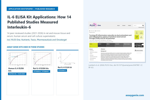
IL-6 ELISA Kit: 14 Studies in Rat, Mouse and Human Samples

Summary
Assay Genie IL-6 sandwich ELISA kits, and one sandwich chemiluminescent immunoassay (CLIA), were used in the 14 studies summarised here. They measured interleukin-6 in rat colon, kidney, brain and heart homogenates, mouse brain and serum, human clinical serum, and supernatants from macrophages, fibroblasts and breast cancer cells. In most, IL-6 was the protein-level inflammation readout used to test whether a treatment, knockdown or disease state changed cytokine output.
Key takeaways
- 14 published studies used Assay Genie IL-6 kits: sandwich ELISAs and one rat IL-6 CLIA (RTES00015).
- Samples were rat colon, kidney, brain and heart homogenates, mouse brain and serum, human clinical serum, and supernatants from macrophages, fibroblasts and breast cancer cells.
- IL-6 was usually measured with TNF-α, IL-1β or both, and several groups used an inhibitor or agonist to test which pathway an IL-6 change depended on.
- Serum IL-6 was in the low pg/ml range in mild cognitive impairment and did not separate haemodialysis patients from controls, so assay sensitivity and pre-analytical control matter most in these settings.
- Five papers gave no catalogue number; reporting catalogue number, species and lot aids reproducibility.
Products cited in these studies
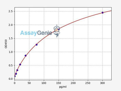
Human IL-6 ELISA Kit (HUFI00180)
Cited by catalogue number in study [7] (Atsopardi et al., 2025).
View kit →
Human IL6 PharmaGenie ELISA Kit (HUDC0061)
Used in study [9] (Katsipis et al., 2024).
View kit →
Rat IL-6 ELISA Kit (RTFI00034)
Listed in the citation record for study [3] (Kattan et al., 2026).
View kit →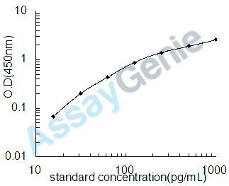
Rat IL-6 ELISA Kit (RTEB0061)
Used in studies [5] and [6] (Alrashdi et al., 2023; AlTamimi et al., 2023, Nutrients).
View kit →
Rat IL-6 CLIA Kit (RTES00015)
Used in study [1] (Akeel et al., 2026) and listed in the citation record for study [2] (Alemam et al., 2026).
View kit →At a glance
| Study | Model / species | Sample type | Product | What it measured | Role in study |
|---|---|---|---|---|---|
| Akeel et al., 2026, Pharmaceuticals [1] | Male Wistar rats, DMH ± morin liposomes | Colon homogenate | Rat IL-6 CLIA Kit (RTES00015) | Colonic IL-6 | Panel with TNF-α, IL-1β |
| Alemam et al., 2026, Reprod Toxicol [2] | Male Sprague-Dawley rats, methotrexate | Not accessible | RTES00015 (citation record) | IL-6 | Inflammation readout |
| Kattan et al., 2026, Med Oncol [3] | Wistar rats, DMH ± honokiol liposomes | Not accessible | Rat IL-6 ELISA Kit (RTFI00034) (citation record) | IL-6 | Raised by DMH |
| AlTamimi et al., 2023, Saudi Pharm J [4] | Male Wistar rats, STZ diabetes ± eriodictyol ± ML385 | Kidney homogenate | Assay Genie IL-6 ELISA; printed cat. no. is another kit | Renal IL-6 | Nrf2-dependent effect |
| Alrashdi et al., 2023, Toxics [5] | Male Wistar rats, PTZ ± rosemary extract | Whole-brain homogenate | Rat IL-6 ELISA Kit (RTEB0061) | Brain IL-6 | Neuroinflammation endpoint |
| AlTamimi et al., 2023, Nutrients [6] | Male Wistar rats, STZ ± AKBA ± compound C | Left-ventricle homogenate | RTEB0061 | Cardiac IL-6 | AMPK-dependent effect |
| Atsopardi et al., 2025, J Neurosci Res [7] | Male C57BL/6 mice, acrylamide ± CBD | Three brain-region homogenates | Cited as HUFI00180 (Human IL-6 ELISA Kit) | IL-6, pg/mg protein | Region-specific effect |
| Huang et al., 2021, eCAM [8] | Mouse BMDMs + oxLDL; ApoE⁻/⁻ mice | Supernatant; serum | ELISA Genie (no cat. no.) | Secreted, serum IL-6 | GDF-15 lowered IL-6 |
| Katsipis et al., 2024, Neurosci Appl [9] | 74 adults with MCI | Fasting serum | Human IL-6 PharmaGenie ELISA Kit (HUDC0061) | Serum IL-6 | Fell with exercise |
| Saboor et al., 2025, J King Saud Univ Sci [10] | 42 haemodialysis patients, 42 controls | Serum | Assay Genie (no cat. no.) | Serum IL-6 | No significant difference |
| Nwanguma et al., 2025, Int J Clin Case Rep Investig [11] | 156 hypertensive, 52 normotensive adults | Fasting serum | Printed “HUF100180” | Serum IL-6 | Higher in hypertension |
| Lambert et al., 2026, PLoS One [12] | THP-1 macrophages + LPS | Supernatant | Assay Genie human IL-6 (no cat. no.) | Secreted IL-6 | Extract lowered IL-6 |
| Ortiz-Flores et al., 2026, Aesthetic Plast Surg [13] | Human fibroblasts; J774A.1 macrophages | Supernatant, 24–72 h | Assay Genie (no cat. no.) | Secreted IL-6 | Filler raised IL-6 |
| Morris et al., 2021, Oncotarget [14] | MDA-MB-231 cells, eIF3e siRNA | Conditioned medium | ELISA Genie (no cat. no.) | Secreted IL-6 | Senescence secretome |
Why measure IL-6?
IL-6 is a pleiotropic cytokine made by macrophages, fibroblasts, endothelial and other cells in response to Toll-like receptor ligands, IL-1β and TNF-α, largely through NF-κB. It drives the hepatic acute-phase response, including CRP and hepcidin, shapes lymphocyte responses and is released by contracting muscle as a myokine.
IL-6 signals through the IL-6 receptor α-chain (IL-6R) and the shared transducer gp130 (IL6ST). In classic signalling, IL-6 binds membrane IL-6R, found mainly on hepatocytes and some leukocytes. In trans-signalling, IL-6 binds soluble IL-6R, and because gp130 is expressed almost everywhere, the complex can activate cells lacking membrane IL-6R. Both routes activate JAK1 (with JAK2 and TYK2), which phosphorylates STAT3. STAT3 dimers enter the nucleus and induce target genes, including the feedback inhibitor SOCS3.
Because IL-6 is secreted, a sandwich immunoassay of serum, medium or tissue extract gives a protein-level readout of inflammatory output that mRNA data cannot.

How the product was used
Rat tissue homogenates
Akeel et al. (2026) compared free and liposomal morin in male Wistar rats given 1,2-dimethylhydrazine (DMH) weekly for 10 weeks. Colon homogenates (10% w/v, cold phosphate buffer) were assayed for TNF-α, IL-6 and IL-1β with kits listed “respectively” as AEES00516, RTES00015 and AEES02613, confirming the Rat IL-6 CLIA kit as the IL-6 assay. Values were protein-normalised. DMH markedly raised colonic IL-6, and both morin forms lowered it, although it stayed modestly above control. Alongside the TNF-α, IL-1β and MPO data, this supported the anti-inflammatory conclusion [1].
Alemam et al. (2026) compared icariin-loaded nanoliposomes with crude icariin in 60 male Sprague-Dawley rats with methotrexate-induced testicular toxicity. The abstract reports NF-κB-mediated inflammation that treatment suppressed, but it does not mention IL-6. The citation record lists RTES00015 for IL-6. Sample type and IL-6 result: methods detail not accessible [2].
Kattan et al. (2026), with authors shared with [1], used a similar 10-week DMH design in 60 Wistar rats to compare liposomal and free honokiol. The abstract reports that DMH increased inflammatory mediators, including IL-6, and that the liposomal form suppressed inflammatory signalling more strongly. The citation record lists RTFI00034. Methods detail not accessible [3].
AlTamimi et al. (2023, Saudi Pharm J) gave eriodictyol (20 mg/kg orally, 12 weeks) to streptozotocin (STZ)-diabetic male Wistar rats, with or without the Nrf2 inhibitor ML385. Diabetes raised kidney-homogenate IL-6 and eriodictyol lowered it. ML385 abolished the reduction, so the authors concluded that the effect depends on Nrf2. The IL-6 kit is printed as “RTEB1811”. In Assay Genie’s catalogue, that is a rat glutathione ELISA, and the number reappears in the paper’s oxidative-marker list. The exact IL-6 kit cannot be confirmed [4].
Alrashdi et al. (2023) gave Rosmarinus officinalis extract (100 mg/kg) to male Wistar rats with pentylenetetrazol (PTZ)-induced epilepsy, alongside a valproate-treated group. Whole-brain homogenates were assayed with Assay Genie kits; by position in the analyte list, RTEB0061 was the IL-6 kit. PTZ raised brain IL-6. Both treatments lowered it, with the extract below valproate but still above control [5].
AlTamimi et al. (2023, Nutrients) gave 11-keto-β-boswellic acid (AKBA in the paper; 10 mg/kg orally, 8 weeks) to STZ-diabetic male Wistar rats, with or without the AMPK inhibitor compound C. Left-ventricle IL-6 (Cat# RTEB0061) rose with diabetes and fell with AKBA, and compound C blunted the effect. The authors interpreted this as AMPK-dependent anti-inflammatory activity [6].
Mouse brain, macrophages and serum
Atsopardi et al. (2025) gave male C57BL/6 mice acrylamide (10 mg/kg i.p., 5 days), cannabidiol (CBD, 10 mg/kg i.p., 10 days) or both. IL-6 in hippocampus, cortex and striatum homogenates was measured with a kit cited as HUFI00180 and reported as pg/mg protein. Acrylamide raised IL-6 in all three regions. CBD lowered it in cortex and striatum, not hippocampus. HUFI00180 is Assay Genie’s Human IL-6 ELISA Kit, and the paper does not discuss mouse cross-reactivity [7].
Huang et al. (2021) exposed mouse bone-marrow-derived macrophages to oxidised LDL (50 µg/ml, 48 h) ± recombinant GDF-15, and treated high-fat-fed ApoE⁻/⁻ mice with GDF-15. IL-6 in supernatant and serum was measured with ELISA Genie kits (no catalogue number). oxLDL raised secreted IL-6 and GDF-15 suppressed it; serum IL-6 was also lower. A TLR4 agonist reversed the suppression, linking the effect to TLR4 [8].
Human clinical serum
Katsipis et al. (2024) followed 74 people with mild cognitive impairment in Greece for 3 months: no intervention, exercise, or exercise plus cognitive training. Fasting serum at baseline and 3 months was assayed in duplicate with the Human IL-6 PharmaGenie ELISA (#HUDC0061). Baseline means were about 4 pg/ml. IL-6 rose in controls and fell in both intervention groups [9].
Saboor et al. (2025) compared 42 adults on haemodialysis with 42 healthy controls in Sharjah, UAE, to examine FGL1 and hepcidin in the anaemia of chronic kidney disease. Serum stored at −80 °C was assayed for FGL1, erythroferrone, hepcidin and IL-6 with Assay Genie ELISAs (no catalogue numbers). IL-6 did not differ significantly between groups, and its correlation with hepcidin was weak and non-significant. The authors cite heterogeneous inflammation, a single time point, pre-analytical variability and sample size [10].
Nwanguma et al. (2025) recruited hypertensive adults (104 treated, 52 untreated) and 52 normotensive controls in Owerri, Nigeria. Fasting serum was stored at −20 °C and assayed with an Assay Genie IL-6 ELISA whose catalogue number is printed as “HUF100180”, which is not a current Assay Genie number. IL-6 was higher in treated hypertensives than in controls, lower than in untreated hypertensives, and correlated with systolic pressure [11].
Cell culture supernatants
Lambert et al. (2026) stimulated PMA-differentiated THP-1 macrophages with LPS (250 ng/ml, 24 h) alongside chestnut wood extract, urolithin B, quercetin, rosiglitazone or a PPARγ antagonist. Supernatant IL-6 was measured with the “Assay Genie Human IL-6 ELISA Kit” (no catalogue number). The extract reduced IL-6, and the reduction persisted under PPARγ antagonism. The authors therefore proposed a PPARγ-independent route [12].
Ortiz-Flores et al. (2026) exposed normal human dermal fibroblasts and J774A.1 macrophages (a murine line), alone and in Transwell co-culture, to extracts of Hylan Gel Dermal Filler DX and comparator fillers. Supernatants were taken at 24, 48 and 72 h and run in triplicate with Assay Genie ELISAs (no catalogue numbers). The filler raised IL-6 in both cell types at 48 and 72 h. The authors suggest that this may support collagen synthesis but did not test causation [13].
Morris et al. (2021) silenced eIF3e in MDA-MB-231 breast cancer cells. They collected 24-h serum-free conditioned medium, clarified it and adjusted it to equal cell numbers before assay with an ELISA Genie kit. Knockdown increased secreted IL-6 about tenfold. Rapamycin roughly halved the rise in IL-6 transcripts. Together, these data supported an mTORC1-linked senescence-associated secretory phenotype [14].
Cross-study insights
- Sample types. Tissue homogenates were most common [1, 4–7], followed by serum [8–11] and culture supernatants [8, 12–14].
- Normalisation. Homogenate IL-6 was protein-normalised [1, 7]. Conditioned medium was collected serum-free and adjusted to cell number [14]. Clinical studies reported fasting collection [9, 11] and frozen storage [10, 11].
- IL-6 as a mechanistic endpoint. Groups tested pathway dependence with ML385 (Nrf2) [4], compound C (AMPK) [6], a PPARγ antagonist [12], a TLR4 agonist [8] and rapamycin (mRNA only) [14].
- Panels. IL-6 was usually measured with TNF-α, IL-1β or both [1, 3–7, 11–13].
- Low and null signals. Serum IL-6 in mild cognitive impairment was in the low pg/ml range [9], and IL-6 did not separate haemodialysis patients from controls [10]. Assay sensitivity and pre-analytical control matter most in these settings.
- Catalogue reporting. Five papers gave no catalogue number [8, 10, 12–14]. Two printed numbers that do not identify an IL-6 kit [4, 11], and one cited a human kit for mouse tissue [7]. In the same group’s Nutrients paper, RTEB1811 is listed for GSH [6]. Reporting catalogue number, species and lot aids reproducibility.

Practical considerations
Generic best practice and product-page information, not reported by the cited studies:
- Match the species. Human, mouse and rat IL-6 differ in sequence. Use a species-matched kit, or confirm cross-reactivity on the datasheet first.
- ELISA or CLIA. The RTES00015 product page describes a sandwich CLIA with biotinylated detection antibody and avidin-HRP, read as relative light units on a chemiluminescence analyser. Colorimetric ELISAs need an absorbance reader. Check each product page for range and sensitivity.
- Serum and plasma. Standardise clotting, centrifugation and collection time, since exercise and time of day can shift circulating IL-6. Aliquot samples and avoid repeated freeze–thaw.
- Tissue. Homogenise in cold buffer with protease inhibitors, clarify, measure total protein and report IL-6 per mg protein. Pilot dilutions to stay within the standard curve.
- Conditioned medium. Use a defined, preferably serum-free, collection window, a medium-only blank and cell-number normalisation.
- Controls. Run standards and samples in duplicate, and include a stimulated positive control (e.g. LPS-treated macrophage supernatant).
Measuring IL-6?
The human and rat IL-6 ELISA and CLIA kits cited in these 14 studies are available from Assay Genie, with expert technical support.
View IL-6 research products →Frequently asked questions
What samples can an IL-6 ELISA kit measure?
Sandwich IL-6 ELISAs are widely used with serum, plasma, cell culture supernatant and tissue homogenates. The studies above used rat colon, kidney, brain and heart homogenates, mouse brain and serum, human serum, and supernatants from macrophages, fibroblasts and cancer cells. Check each product page for validated sample types and volumes.
What is the difference between an IL-6 ELISA and an IL-6 CLIA?
Both are sandwich immunoassays using a capture antibody and a labelled detection antibody. An ELISA generates a coloured product read by absorbance. A CLIA generates light, measured as relative light units, so it needs a chemiluminescence-capable reader. Compare range and sensitivity on each product page before choosing.
Can I use a human IL-6 ELISA kit on mouse or rat samples?
Only if the datasheet confirms cross-reactivity. Human, mouse and rat IL-6 differ in sequence, and antibody pairs are usually species-specific. One study above cited a human-kit catalogue number for mouse brain without discussing cross-reactivity. For new work, a species-matched rat or mouse IL-6 kit is the safer choice.
How should IL-6 in tissue homogenate be normalised?
Measure total protein, for example by BCA, in the same clarified homogenate and report IL-6 per mg protein, as two of the cited studies did. Keep the buffer, tissue-to-buffer ratio and centrifugation conditions identical across groups, and dilute samples so they fall within the kit’s standard curve.
Why measure IL-6 together with TNF-α and IL-1β?
These cytokines are induced together by NF-κB-driven inflammatory signalling. A panel shows whether a treatment shifts the overall inflammatory response or only one mediator. Most of the studies above paired IL-6 with TNF-α, IL-1β or both, and several added an inhibitor to test which pathway the IL-6 change depended on.
References
- Akeel MA, Elmorsy EM, Alshammari FM, Shaalan AAM, et al. Liposomal morin attenuates DMH-associated colonic oxidative stress, inflammation, and apoptosis/autophagy-related dysregulation in rats. Pharmaceuticals. 2026;19(9):1400. doi:10.3390/ph19091400.
- Alemam A, Jamil S, Hassan H, Elmorsy EM. Protective effects of icariin-loaded nanoliposomes against methotrexate-induced testicular toxicity via modulation of NRF2/NF-κB and ferroptosis signaling. Reproductive Toxicology. 2026;145:109327. doi:10.1016/j.reprotox.2026.109327.
- Kattan SW, Almars AI, Elmorsy EM, Al-Ghafari AB, et al. Liposomal honokiol attenuates dimethylhydrazine-induced colon carcinogenesis in rats through modulation of oxidative stress, inflammation, apoptosis, autophagy, and lipid metabolism pathways. Medical Oncology. 2026;43:215. doi:10.1007/s12032-026-03330-6.
- AlTamimi JZ, AlFaris NA, Alshammari GM, Alagal RI, et al. Protective effect of eriodictyol against hyperglycemia-induced diabetic nephropathy in rats entails antioxidant and anti-inflammatory effects mediated by activating Nrf2. Saudi Pharmaceutical Journal. 2023;31(11):101817. doi:10.1016/j.jsps.2023.101817.
- Alrashdi J, Albasher G, Alanazi MM, Al-Qahtani WS, et al. Effects of Rosmarinus officinalis L. extract on neurobehavioral and neurobiological changes in male rats with pentylenetetrazol-induced epilepsy. Toxics. 2023;11(10):826. doi:10.3390/toxics11100826.
- AlTamimi JZ, AlFaris NA, Alshammari GM, Alagal RI, et al. The protective effect of 11-keto-β-boswellic acid against diabetic cardiomyopathy in rats entails activation of AMPK. Nutrients. 2023;15(7):1660. doi:10.3390/nu15071660.
- Atsopardi K, Mesiakaris K, Sotiropoulos I, Margarity M, Poulas K. Cannabidiol as a neuroprotective agent in acrylamide-induced neurotoxicity: effects on oxidative stress, inflammation, and cholinergic function in male mice. Journal of Neuroscience Research. 2025;103(12):e70098. doi:10.1002/jnr.70098.
- Huang H, Chen Z, Li Y, Gong K, et al. GDF-15 suppresses atherosclerosis by inhibiting oxLDL-induced lipid accumulation and inflammation in macrophages. Evidence-Based Complementary and Alternative Medicine. 2021;2021:6497568. doi:10.1155/2021/6497568.
- Katsipis G, Tzekaki EE, Andreadou EG, Mouzakidis C, et al. The effect of physical exercise with cognitive training on inflammation and Alzheimer’s disease biomarkers of mild cognitive impairment patients. Neuroscience Applied. 2024;3:104085. doi:10.1016/j.nsa.2024.104085.
- Saboor M, Hashim HN, Guella A, Alkhayyal N. Hepatokine FGL1 and hepcidin in anemia of chronic kidney disease: a cross sectional study. Journal of King Saud University – Science. 2025;37. doi:10.25259/JKSUS_1251_2025.
- Nwanguma ED, Nwanjo H, Nwosu D, Nnodim J, Edward U. Evaluation of levels of interleukin 6 (IL-6) and tumour necrosis factor-alpha of hypertensive subjects undergoing care at Federal Teaching, Owerri. International Journal of Clinical Case Reports and Investigations. 2025;2(1):14.
- Lambert D, Buisine M, Billamboz M, Jawhara S. Turning off inflammation naturally via dual antioxidant and anti-inflammatory actions of chestnut wood extract through PPARγ and NF-κB pathways. PLoS One. 2026;21(4):e0347987. doi:10.1371/journal.pone.0347987.
- Ortiz-Flores RM, Escamilla-Sanchez A, Cidoncha-Morcillo B, Mastronardi L, et al. Enhanced facial rejuvenation: biostimulatory effects of Hylan Gel Dermal Filler DX on collagen synthesis and tissue regeneration. Aesthetic Plastic Surgery. 2026;50(2):707–725. doi:10.1007/s00266-025-05245-5.
- Morris C, Durand S, Jalinot P. Decreased expression of the translation factor eIF3e induces senescence in breast cancer cells via suppression of PARP1 and activation of mTORC1. Oncotarget. 2021;12(7):649–664. doi:10.18632/oncotarget.27923.
Recent Posts
-
IL-6 ELISA Kit: 14 Studies in Rat, Mouse and Human Samples
SummaryAssay Genie IL-6 sandwich ELISA kits, and one sandwich chemiluminescent immunoassay (CLIA) …21st Nov 2026 -
Fatty Acid Oxidation Assay Kit: 20 Published Applications
SummaryThe Assay Genie Fatty Acid Oxidation (FAO) Assay Kit (BR00001) is a colorimetric plate-rea …20th Nov 2026 -
Mycoplasma Detection Kits for Cell Culture: A Guide
SummaryA mycoplasma detection kit screens cell-culture supernatant for mycoplasma DNA so contaminate …20th Nov 2026