

MycoGenie Rapid Mycoplasma Detection Kit (MORV0011): Published Uses

Summary
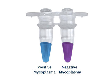
The MycoGenie Rapid Mycoplasma Detection Kit (MORV0011) is a one-hour, single-temperature colour test that screens 1 µl of cell-culture supernatant for mycoplasma: purple means negative and sky blue means positive. Two recent studies, one on endothelin-1 and gasdermin E in ovarian cancer metastasis and one on NNMT in kidney tubular senescence, cite MycoGenie as the mycoplasma check for their human cancer cell lines and mouse renal tubular epithelial cells.
Key takeaways
- Roman et al. (2026) tested their ovarian cancer and other human cell lines with MycoGenie (#MORV0011-50) alongside STR authentication [1].
- Chanvillard et al. (2025, preprint) regularly tested conditionally immortalised mouse renal tubular epithelial cells with the MycoGenie rapid kit [2].
- In both papers mycoplasma testing was a quality-control precondition; neither reports test results or sampling details.
- The kit runs 60 min at 60 °C on 1 µl of supernatant with a visual purple-to-sky-blue read-out (product page) [7].
Product cited in these studies
MycoGenie Rapid Mycoplasma Detection Kit (MORV0011)
Used by Roman et al. (2026) and Chanvillard et al. (2025) for mycoplasma quality control of human cancer cell lines and mouse renal tubular epithelial cells.
View kit →At a glance
| Study | Model / species | Sample type | Product | What it measured | Key role in study |
|---|---|---|---|---|---|
| Roman et al., 2026, Cell Death & Disease [1] | Human high-grade serous ovarian carcinoma cells (patient-derived PMOV10, OVCA433, CAOV3, Kuramochi), plus MDA-MB-231 and SW620 | Cultured cells (sample handling not reported) | MycoGenie kit, MORV0011 (#MORV0011-50) | Mycoplasma infection status | Cell-line QC alongside STR authentication |
| Chanvillard et al., 2025, bioRxiv preprint [2] | Conditionally immortalised mouse renal tubular epithelial cells (TECs) from Immorto mice | Cultured cells (sample handling not reported) | Rapid Mycoplasma Detection Kit (MycoGenie, Assay Genie), MORV0011 | Mycoplasma contamination status | Regular QC of the TEC cultures |
Why mycoplasma testing matters
Mycoplasmas are very small bacteria that lack a cell wall. Drexler and Uphoff describe mycoplasma contamination as a major problem in cell culture, with “a virtually unlimited variety of effects” on infected cells, and note that mycoplasmas resist most antibiotics used in culture [3].
Contamination is common. ATCC gives a rate of 15–35% for continuous cell cultures and at least 1% for primary cultures [4]. The DSMZ cell bank reports that almost 20% of cultures it receives from other laboratories are contaminated [5]. A screen of public RNA-seq data found mycoplasma reads in 11% of 884 sequencing series [6]. ATCC lists chromosomal aberrations, disrupted nucleic acid synthesis, altered gene expression, inhibited proliferation and cell death among the effects on host cells [4].
Because these effects reach gene expression, proliferation and viability, an infected line can distort almost any downstream readout, from EMT marker expression to senescence assays. Detecting it requires a dedicated test [4].

How the MycoGenie rapid test works
According to the product page, MycoGenie uses isothermal amplification with a chromogenic buffer, so the reaction runs at one temperature with no thermal cycling, gel, qPCR or ELISA step [7]. The workflow is add, incubate, read:
- Sample. Collect supernatant 3 days after passage or the last media change, at about 90% confluence for adherent lines. Centrifuge suspension cultures at 500 × g for 5 min and use the supernatant.
- Reaction. Add 1 µl of supernatant to the reaction mix (25 µl reaction). Run the kit's positive control and a sterile-water negative control in the same batch.
- Incubate. 60 min at 60 °C in a heated dry-bath, water bath or a PCR instrument used only as a heater.
- Read. Hold the tubes against white paper in bright light: purple is negative, sky blue is positive.

Specifications (from the product page)
| Specification | MycoGenie Rapid Mycoplasma Detection Kit (MORV0011) |
|---|---|
| Method | Isothermal amplification with chromogenic buffer, visual read-out |
| Read-out | Purple (negative) to sky blue (positive) |
| Time | 60 min at 60 °C, single step |
| Sample | 1 µl culture supernatant in a 25 µl reaction |
| Species detected | 28, including the 8 common cell-culture contaminants |
| Sensitivity stated | 500 cfu/µl of supernatant (about 3 × 10² copies) |
| Controls | Positive control included; sterile water as negative control |
| Equipment | Heated dry-bath, water bath or PCR instrument (heating only) |
| Pack sizes | 20 or 50 tests |
| Storage | −20 °C |
The product page also states that the chromogenic buffer tolerates culture-medium inhibitors better than PCR. It lists validation on suspension and adherent lines including CHO, HEK293, Vero, hybridomas and Sf9, and in media such as DMEM, RPMI 1640 and F12 with fetal bovine serum [7]. Check the product page and datasheet for the current protocol before use.
How MycoGenie was used in published research
In both papers, mycoplasma testing is a single quality-control statement in the Methods. Neither paper reports the test results or the sampling details, and only one hints at frequency (“regularly”).
Roman et al. (2026): nuclear gasdermin E in ovarian cancer metastasis
Research question. Roman and colleagues asked whether gasdermin E (GSDME), a pore-forming protein best known for pyroptosis, contributes to metastasis in high-grade serous ovarian carcinoma (HG-SOC) downstream of endothelin-1 (ET-1) signalling [1].
Design and models. The cell panel comprised patient-derived HG-SOC cells (PMOV10, isolated from ascitic fluid), the HG-SOC lines OVCA433, CAOV3 and Kuramochi, MDA-MB-231 breast cancer cells and SW620 colon cancer cells. Cells were grown in RPMI-1640 or DMEM with 10% fetal bovine serum and 1% penicillin-streptomycin. Female nude CD1 mice carried patient-derived or Kuramochi-luciferase intraperitoneal xenografts and were treated with the endothelin receptor antagonist macitentan or vehicle [1].
How MycoGenie was used. Under “Cell culture and reagents”, the authors state that cells were authenticated by short tandem repeat (STR) profiling and “tested for mycoplasma infection, through the MycoGenie kit (Assay Genie, Dublin, Ireland, #MORV0011-50)” [1]. The paper does not say how often cells were tested or give the outcome.
How the readout supported the conclusion. The test was a precondition, not an endpoint. The paper's conclusions rest on EMT and inflammatory readouts in these cell lines. ET-1/ETAR signalling raised GSDME expression through ZEB1. Nuclear GSDME then partnered with ZEB1 to regulate genes such as E-cadherin, vimentin and IL-6, without pyroptosis. Blocking GSDME or the ET-1 receptor reduced EMT and cytokine release [1]. Pairing STR authentication with mycoplasma testing shows that the cell lines behind these readouts were checked for both identity and contamination.
Chanvillard et al. (2025, preprint): NNMT in kidney tubular senescence
This bioRxiv preprint was posted on 6 January 2025 and is not peer reviewed.
Research question. Chanvillard and colleagues asked whether nicotinamide N-methyltransferase (NNMT) drives tubular senescence and fibrosis in chronic kidney disease (CKD) [2].
Design and models. The abstract links NNMT levels to disease severity, senescence markers and fibrosis in patient samples and early-stage mouse models. In vitro, renal tubular epithelial cells (TECs) were isolated from Immorto mice. They were expanded under permissive conditions (33 °C with IFN-γ), then moved to restrictive conditions (37 °C without IFN-γ) for 7 days to downregulate SV40 large T antigen activity before experiments. They were cultured in DMEM/F12 with 10% fetal bovine serum and stimulated with TGF-β. Human iPSC-derived kidney organoids were used separately to test NNMT inhibition [2].
How MycoGenie was used. In the “Renal tubular epithelial cells” methods section, the authors write: “Cells were regularly tested using a Rapid Mycoplasma Detection Kit (MycoGenie, Assay Genie)” [2]. No interval or result is given. The sentence sits under the TEC heading, so this note ties it to the TECs only, not to the organoids. The preprint does not print a catalogue number. MORV0011 is the only Assay Genie product sold under that name.
How the readout supported the conclusion. Again, the test was a QC precondition. According to the abstract, NNMT overexpression in TGF-β-stimulated TECs promoted senescence and a partial epithelial-to-mesenchymal transition, while NNMT inhibition was protective in kidney cells and organoids [2]. Because mycoplasma can alter gene expression and proliferation in host cells [4], regular testing helps protect senescence and EMT readouts in the TEC cultures. A peer-reviewed version later appeared in Cell Reports (2026) under a revised title [8]. This note describes the preprint, which is the version recorded in the citation sheet.
Cross-study insights
- Reporting is minimal. Both papers name the kit and supplier, and only Roman et al. gives the catalogue number (MORV0011-50). Neither gives results or sampling details, and only Chanvillard et al. indicates frequency (“regularly”).
- Different cell types, same check. MycoGenie was used on human cancer lines (including patient-derived ascites cells) and on conditionally immortalised mouse epithelial cells.
- Identity and contamination together. Roman et al. combined STR profiling with mycoplasma testing, so both identity and contamination were checked.
- Antibiotics are no substitute. Roman et al. cultured cells in penicillin-streptomycin. Mycoplasmas lack a cell wall and resist most culture antibiotics [3], so antibiotic-containing medium does not remove the need to test.
Practical protocol considerations
Generic best practice, not from the cited papers:
- Quarantine and test on arrival. Keep new lines apart until they test negative. ATCC and DSMZ both recommend quarantining incoming cultures [4, 5].
- Set a fixed interval. ATCC recommends routine testing but gives no interval [4], so write one into your lab SOP. Also test before banking, after thawing, before costly experiments and before sharing a line.
- Sample at the right time. Follow the kit's sampling point (3 days after passage or media change, about 90% confluence) so that mycoplasma in the supernatant is at its highest [7].
- Run both controls every time. Read each sample against the kit's positive control and the water negative control from the same run.
- Report fully. In your Methods, give the kit name, catalogue number, testing frequency and the result (e.g. all lines negative).
- If a culture is positive. DSMZ normally discards positive lines and replaces them with clean stock, and treats a line only when no clean stock exists. After treatment, DSMZ keeps cells antibiotic-free for at least two weeks before retesting [5].
Testing your cultures for mycoplasma?
The MycoGenie Rapid Mycoplasma Detection Kit cited in these studies is available from Assay Genie, with expert technical support.
View the MycoGenie kit →Frequently asked questions
How long does the MycoGenie mycoplasma test take?
One 60-minute incubation at 60 °C, then the tubes are read by eye. A heated dry-bath or water bath is enough, and a PCR instrument can be used purely as a heater. There is no gel, qPCR or ELISA step, so the result is ready about an hour after the reaction is set up.
What sample does the MycoGenie kit need?
Only 1 µl of cell-culture supernatant, added to a 25 µl reaction. Collect it 3 days after passaging or the last media change, at about 90% confluence for adherent cells. For suspension cultures, centrifuge at 500 × g for 5 minutes and test the cleared supernatant.
How do I read a MycoGenie result?
After incubation, hold the tubes against white paper in bright light. A purple solution is mycoplasma-negative and a sky-blue solution is mycoplasma-positive. Always compare samples with the kit's positive control and a sterile-water negative control run in the same batch.
How sensitive is the MycoGenie Rapid Mycoplasma Detection Kit?
The product page states a sensitivity of 500 cfu of mycoplasma per µl of supernatant, about 3 × 10² copies. It detects 28 mycoplasma species, including the eight most common cell-culture contaminants. For validation data, see the kit's product page and datasheet.
Do antibiotics in culture medium prevent mycoplasma contamination?
No. Mycoplasmas have no cell wall and resist most antibiotics used in cell culture, so penicillin-streptomycin in the medium cannot be relied on to protect against them. Routine testing, quarantine of new lines and good aseptic technique are the main safeguards.
References
- Roman C, Caprara V, Tocci P, Sacconi A, Blandino G, Bagnato A, Sestito R. Nuclear gasdermin E drives endothelin-1-induced metastatic progression independently of the pyroptosis. Cell Death & Disease. 2026;17:45. doi:10.1038/s41419-025-08202-x.
- Chanvillard L, Lantermans H, Wall C, Thevenet J, et al. NNMT promotes tubular senescence and fibrosis in chronic kidney disease. bioRxiv (preprint, not peer reviewed). 2025. doi:10.1101/2025.01.06.631437.
- Drexler HG, Uphoff CC. Mycoplasma contamination of cell cultures: Incidence, sources, effects, detection, elimination, prevention. Cytotechnology. 2002;39:75–90. doi:10.1023/A:1022913015916.
- ATCC. Mycoplasma contamination.
- DSMZ. Mycoplasma detection and eradication.
- Olarerin-George AO, Hogenesch JB. Assessing the prevalence of mycoplasma contamination in cell culture via a survey of NCBI's RNA-seq archive. Nucleic Acids Research. 2015;43:2535–2542. doi:10.1093/nar/gkv136.
- Assay Genie. MycoGenie Rapid Mycoplasma Detection Kit (MORV0011), product page.
- Chanvillard L, et al. NNMT inhibition counteracts tubular senescence and fibrosis in early stages of chronic kidney disease. Cell Reports. 2026;45(1). doi:10.1016/j.celrep.2025.116823.
Recent Posts
-
MycoGenie Rapid Mycoplasma Detection Kit (MORV0011): Published Uses
SummaryThe MycoGenie Rapid Mycoplasma Detection Kit (MORV0011) is a one-hour, single-temperature …22nd Nov 2026 -
Mouse Kynurenine ELISA Kit (MOFI01315): Published Uses
SummaryAssay Genie's Mouse KYN (Kynurenine) ELISA Kit (MOFI01315) is a competitive ELISA that has …22nd Nov 2026 -
IL-6 ELISA Kit: 14 Studies in Rat, Mouse and Human Samples
SummaryAssay Genie IL-6 sandwich ELISA kits, and one sandwich chemiluminescent immunoassay (CLIA) …21st Nov 2026